Photoacoustic imaging converts a brief optical pulse into a detectable sound wave. Light provides selective contrast, while ultrasound carries information from tissue depths that optical imaging alone can struggle to reach.
Ultrasound turns returning sound into a useful map, but brightness depends on how waves meet tissue and how scanners display the result. Understanding those steps clarifies both the image’s power and its limits.
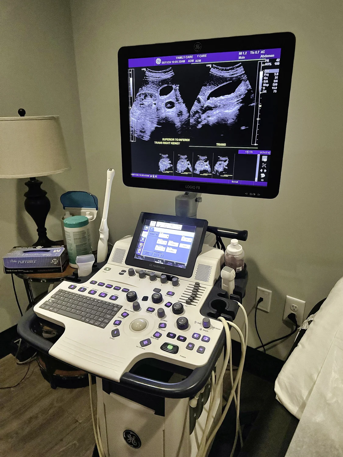
Ultrasound turns echo timing into distance by sending sound pulses and measuring their return. Knowing the speed of sound in tissue lets a scanner place reflecting boundaries at estimated depths.
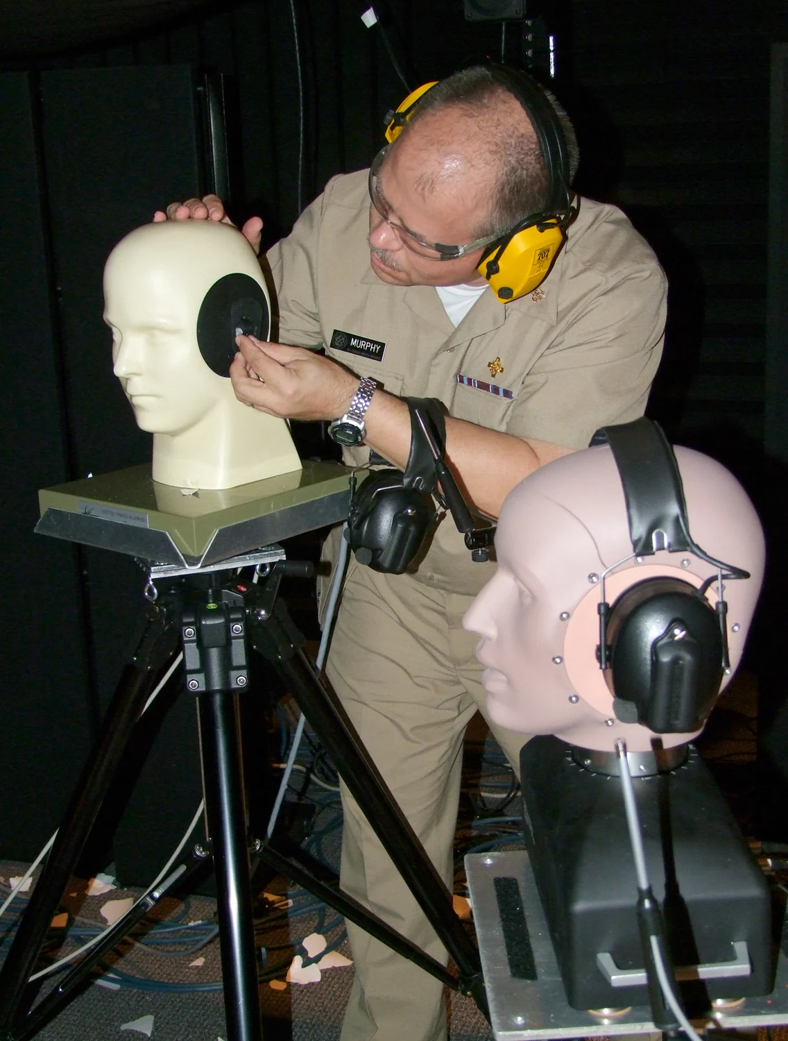
Sound level describes intensity, while exposure duration determines how long the inner ear absorbs that energy. Together with distance and sound character, these factors help explain why a brief impulse and prolonged noise pose different hearing risks.
Elastography turns tissue motion into a map of relative stiffness. Following how a wave travels reveals why this measurement can add information to an anatomical image, while still requiring careful interpretation.
Ultrasound speckle emerges when many echoes combine, so its grainy texture reflects the physics of sound scattering as well as anatomy. Understanding that texture helps explain both image detail and its limits.
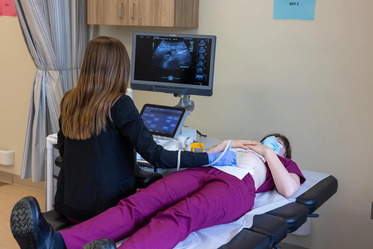
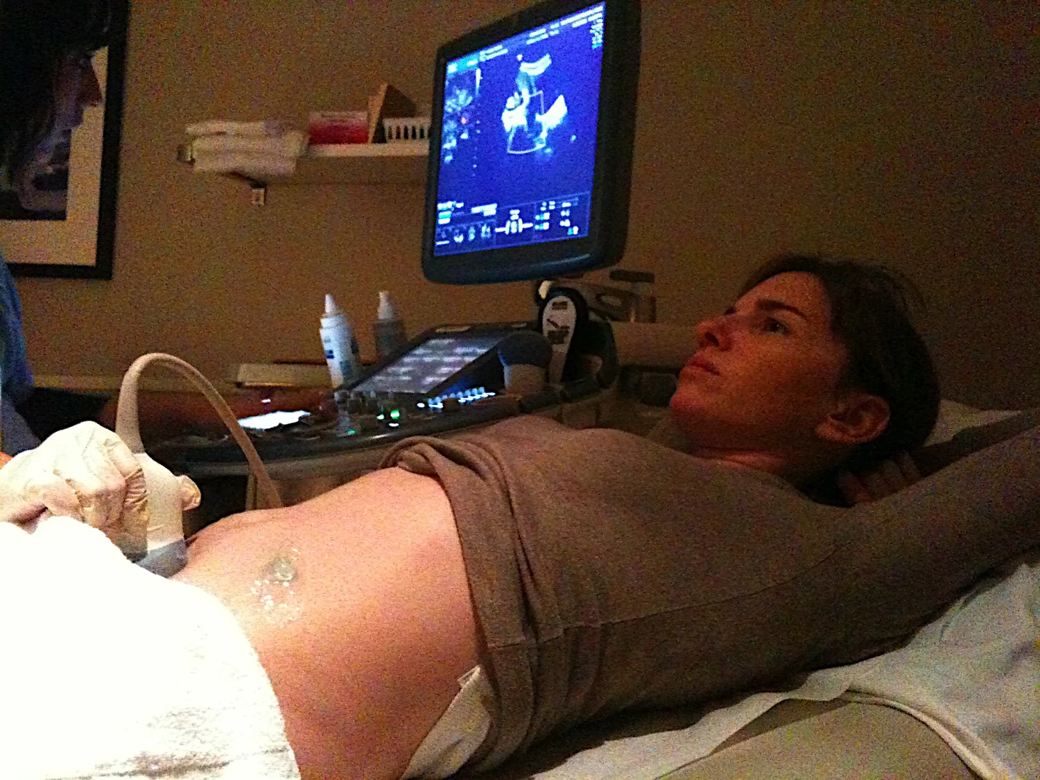
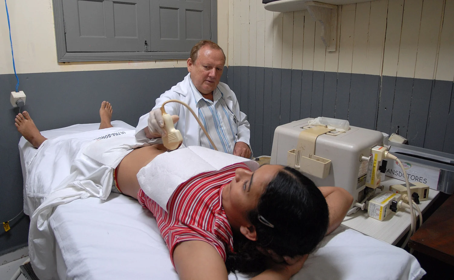
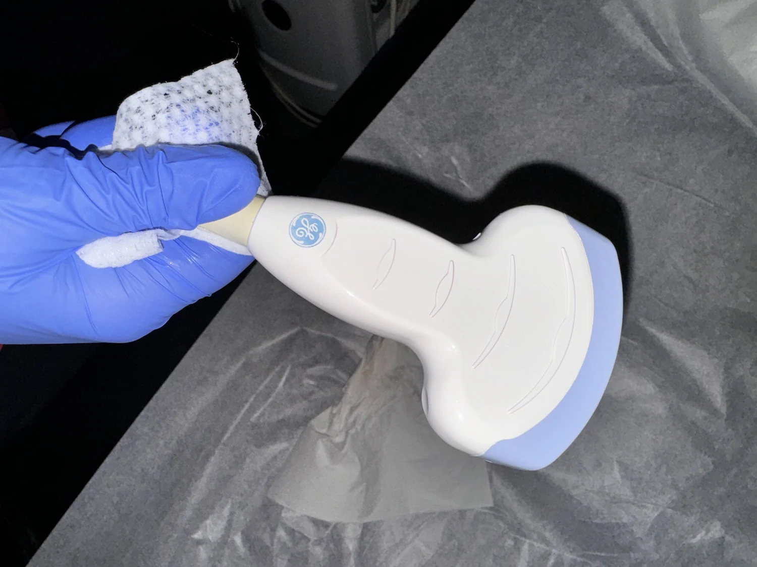
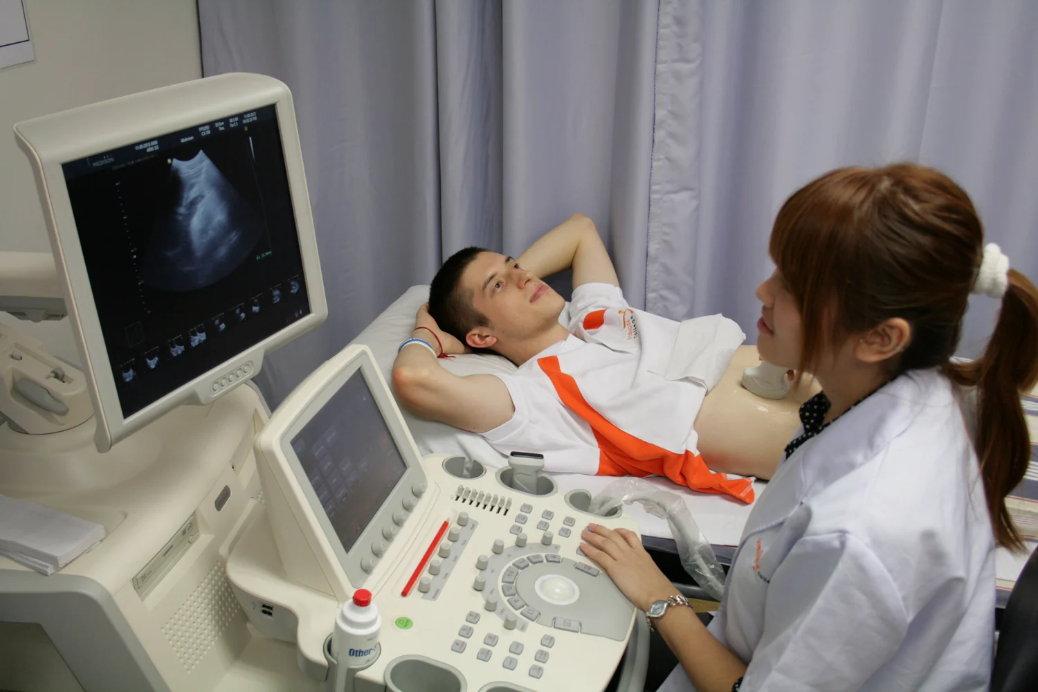
Ultrasound gel removes the air gap between probe and skin so sound can enter the body efficiently. Echo timing and strength then help a scanner map boundaries between tissues in real time.
Decibels compress a vast range of sound pressures into a manageable scale. Understanding the logarithm behind the number helps explain why a small numerical increase can mean a large physical change—and why exposure time matters to hearing.
Diagnostic ultrasound sends sound pulses and listens for echoes to build images. Shock wave lithotripsy uses focused pressure waves to fragment kidney stones, turning acoustic energy toward a therapeutic task.
A 3D ultrasound display is assembled from sound echoes sampled across space. Transducer position, timing, and computer processing turn those measurements into a volume, whose appearance depends on the data collected.
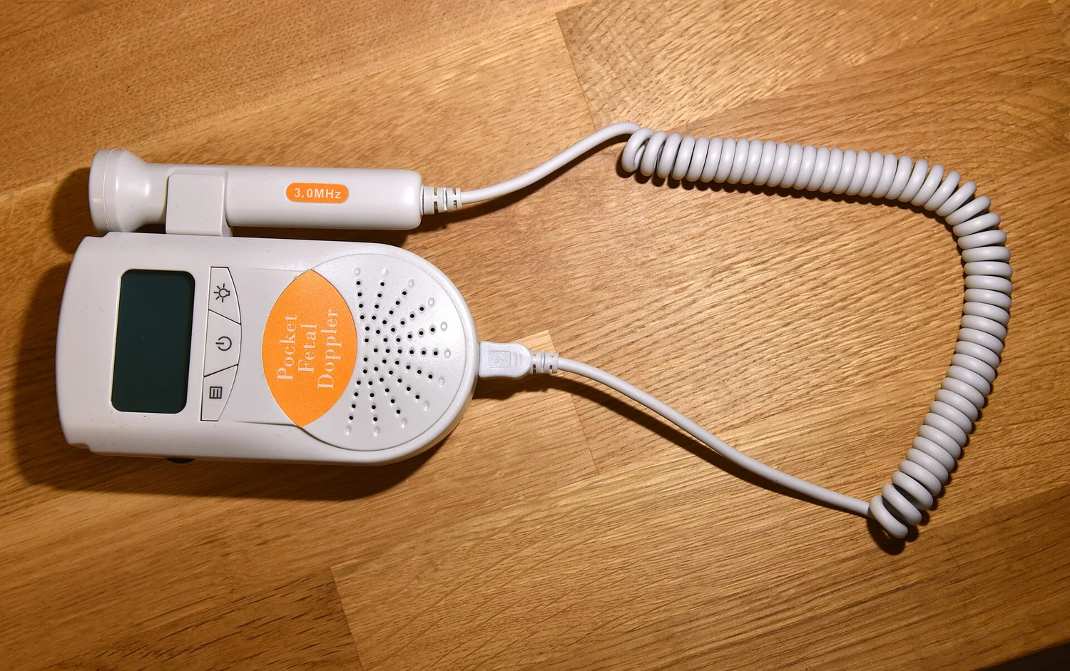
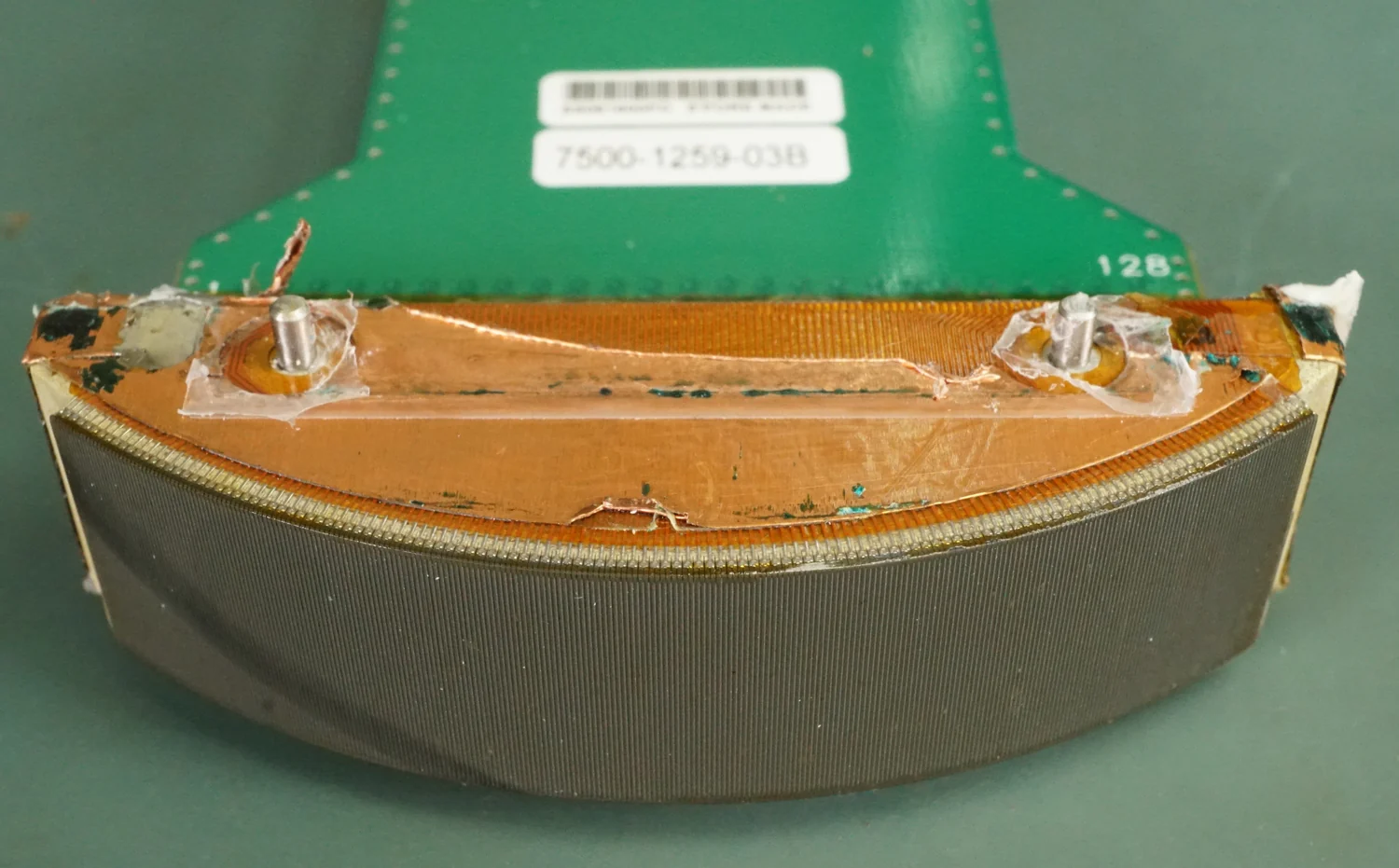
A probe turns electrical pulses into sound and returning echoes back into electrical signals. Their travel times and strengths help a scanner map tissue boundaries, while gel keeps air from blocking the path between skin and probe.
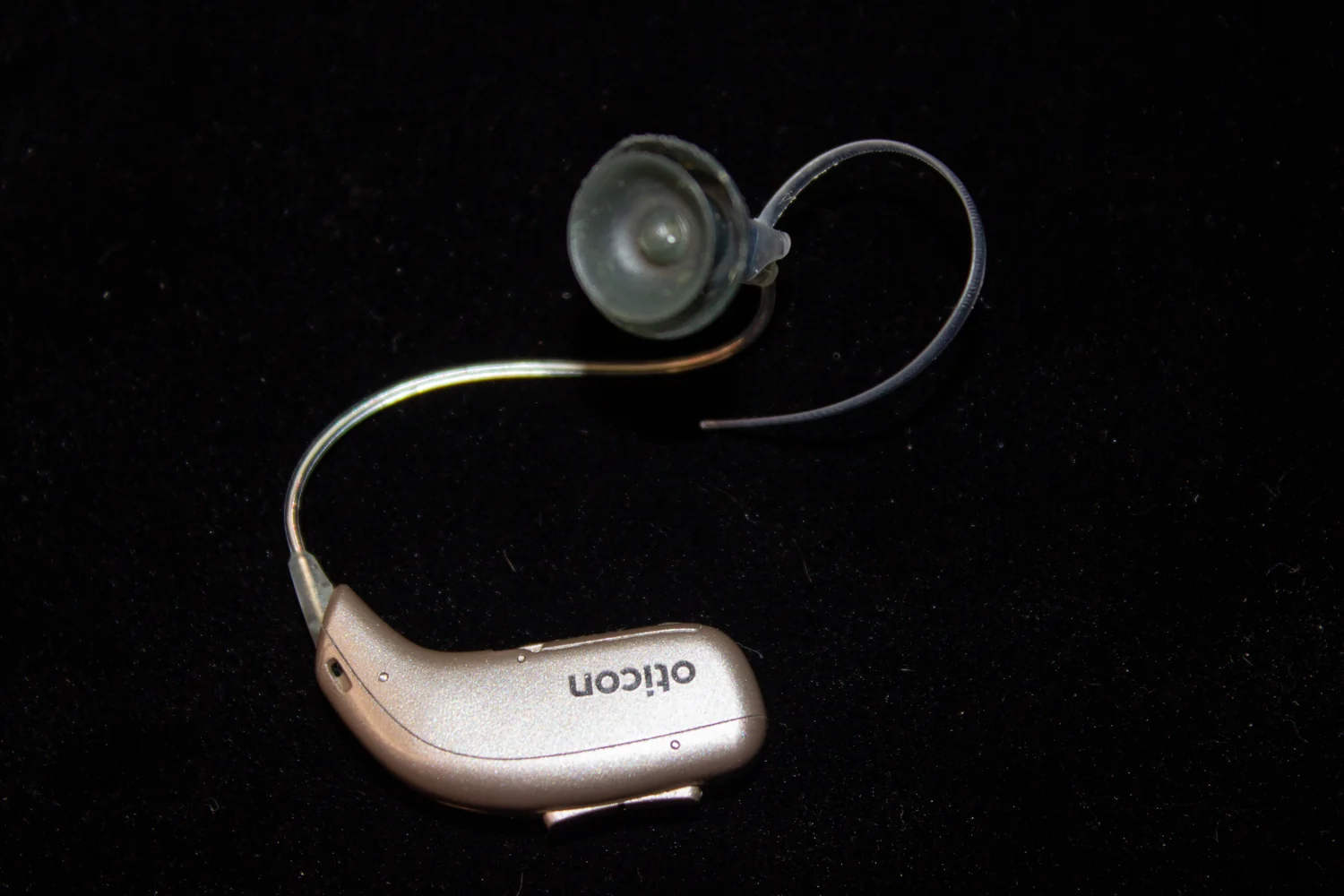
Hearing aids amplify sound vibrations for surviving inner-ear sensory cells to detect. Cochlear implants bypass damaged portions of the ear and electrically stimulate the auditory nerve, creating a different signal the brain must learn to interpret.
Lithotripsy uses focused acoustic pulses to fragment selected kidney stones so pieces can pass through the urinary tract. Stone size, location, and composition shape treatment decisions and alternatives.
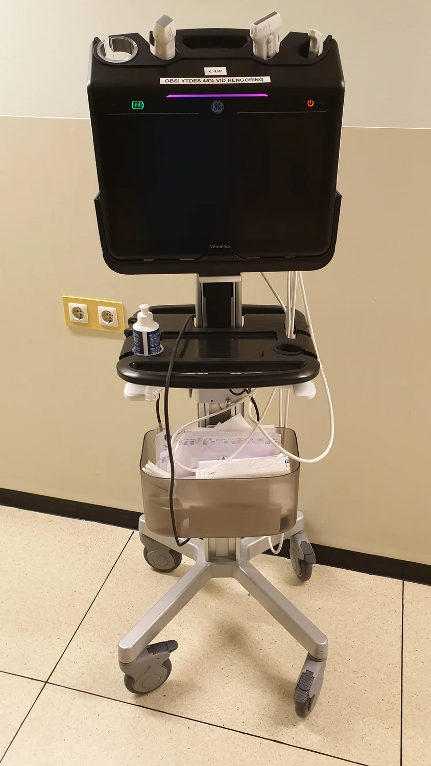
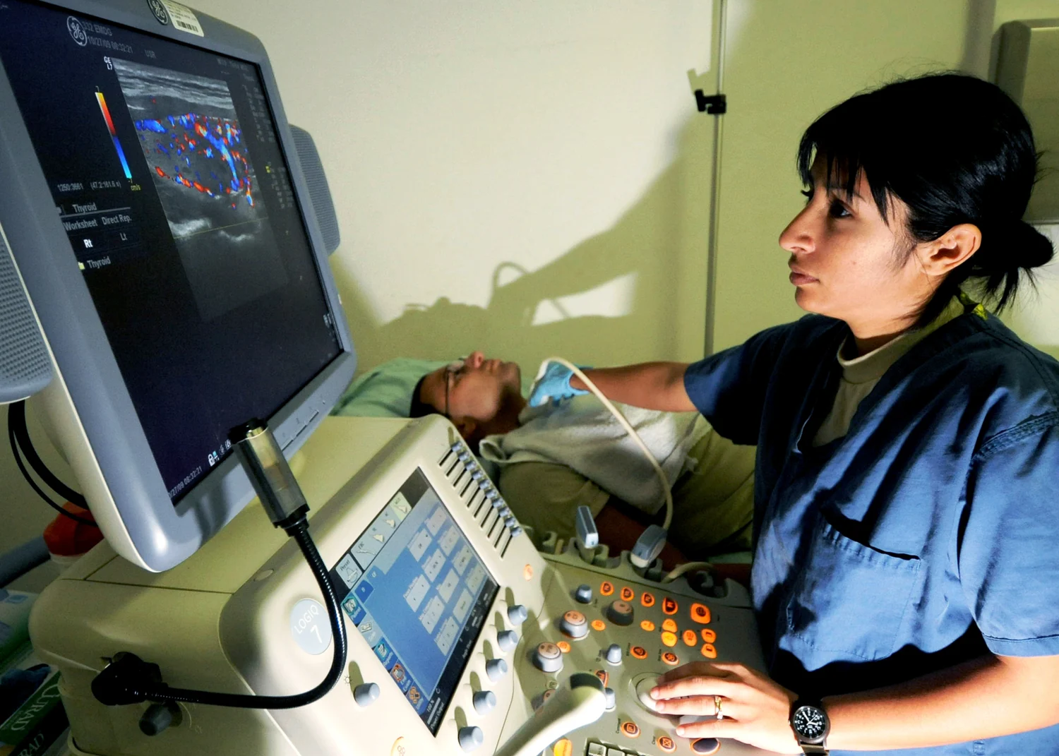
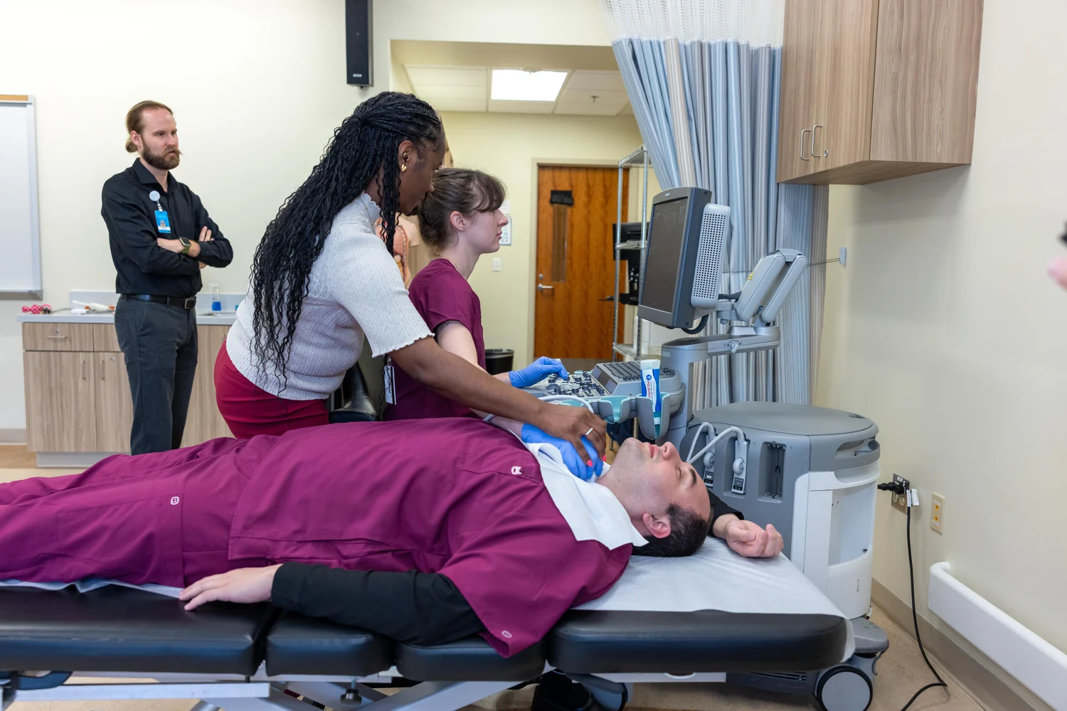
Ultrasound turns echoes into images quickly enough to show organs and blood in motion. That live view can guide procedures, though sound interacts unevenly with bone, air, and soft tissue.
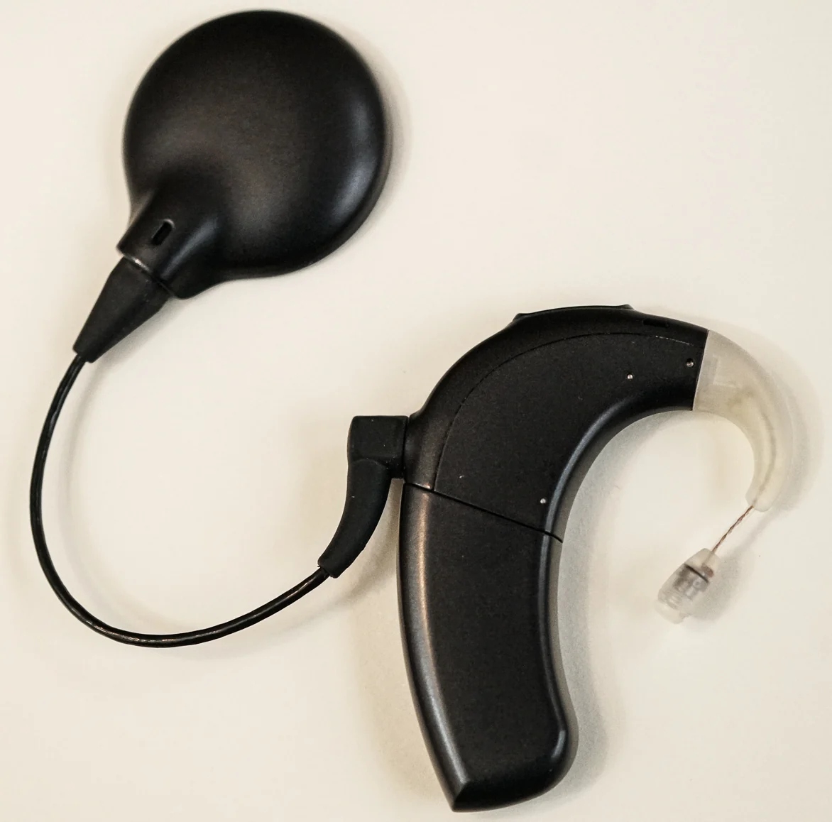
A cochlear implant converts sound into electrical signals and sends them to the auditory nerve, routing around damaged inner-ear hair cells. The result is a learned representation of sound, not normal hearing.
Ultrasonic cleaners use sound-driven cavitation in liquid to help detach debris from intricate instruments. Cleaning removes soil, but it is a distinct step from disinfection and sterilization, which require their own validated processes.
An echocardiogram turns returning sound echoes into moving images of cardiac structures. Different tissues reflect waves differently, and specialized modes can also show how blood moves through chambers and vessels.
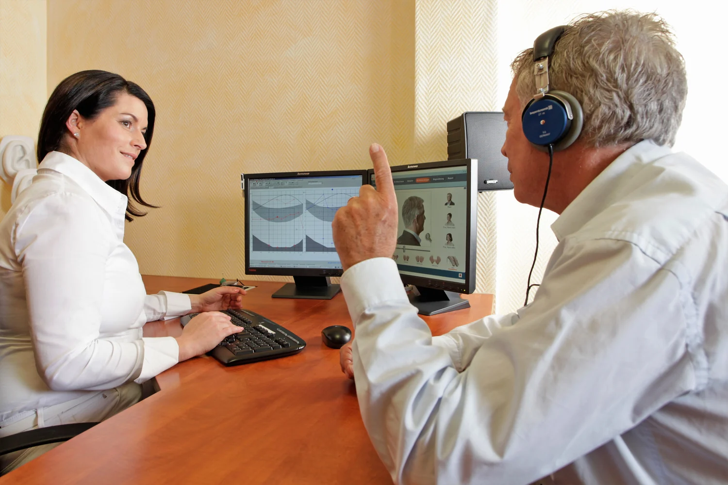
A directional hearing aid can emphasize sounds arriving from a chosen direction, but it cannot perfectly isolate speech from a noisy room. Its usefulness depends on microphone geometry, processing, hearing needs, and the acoustic setting.