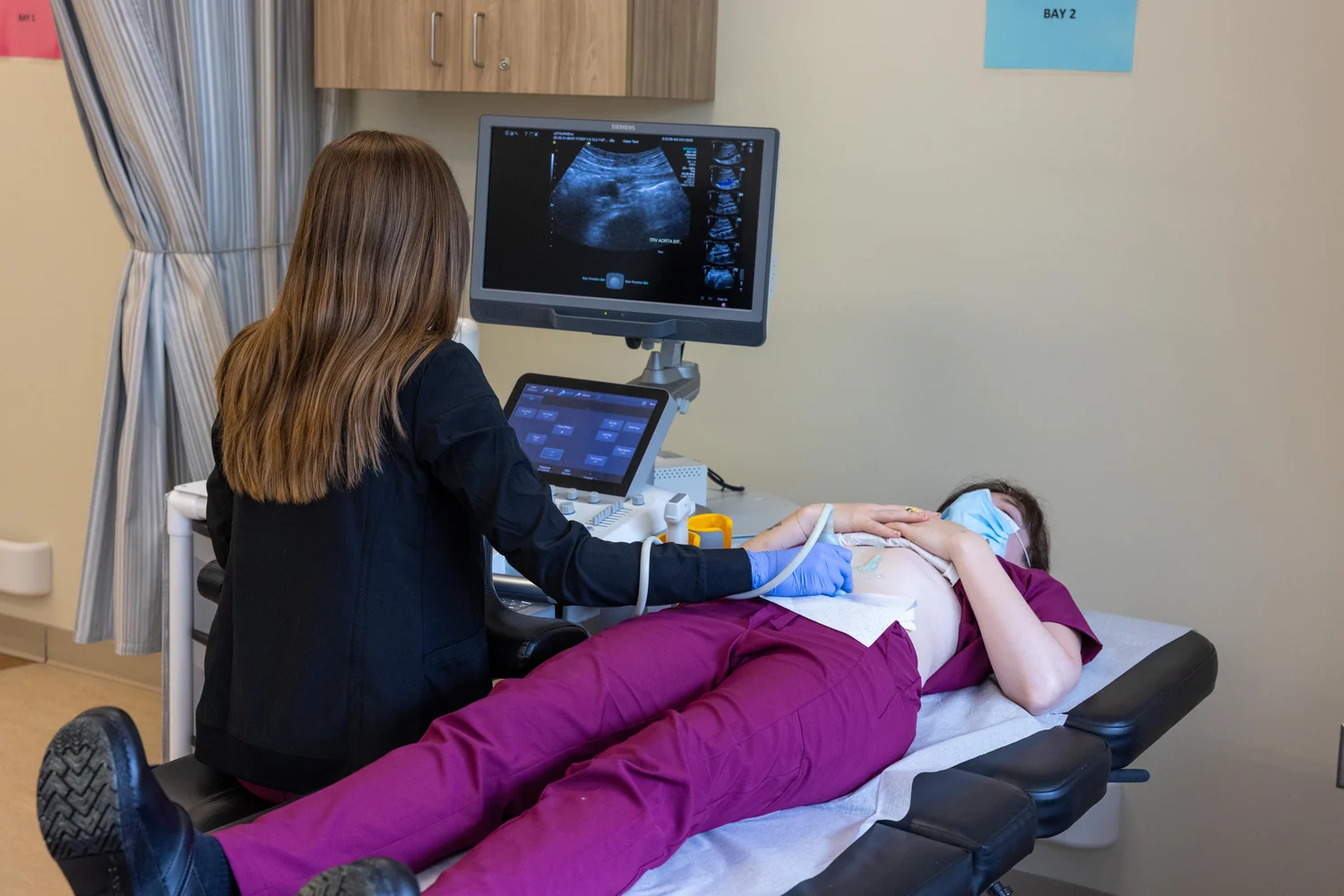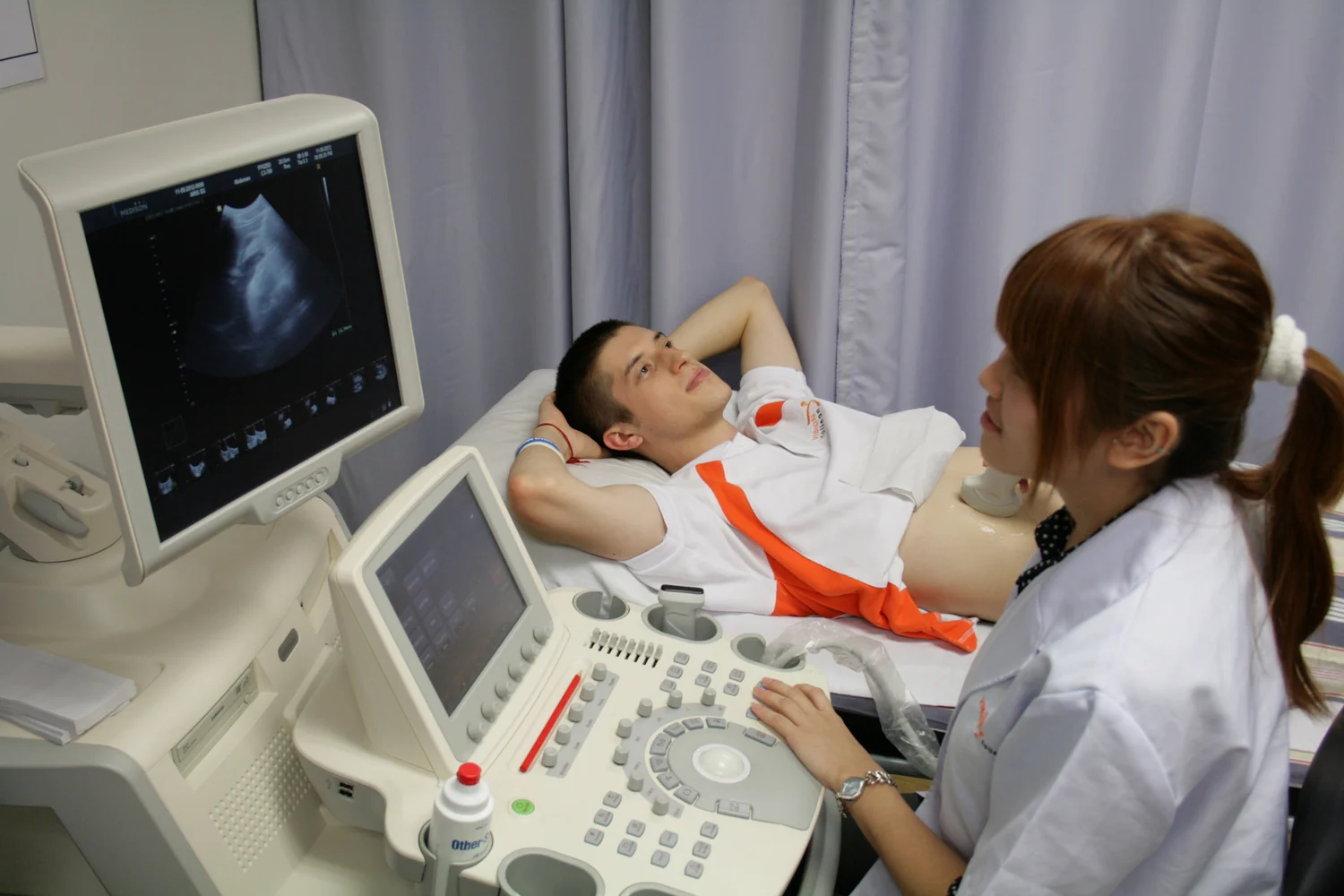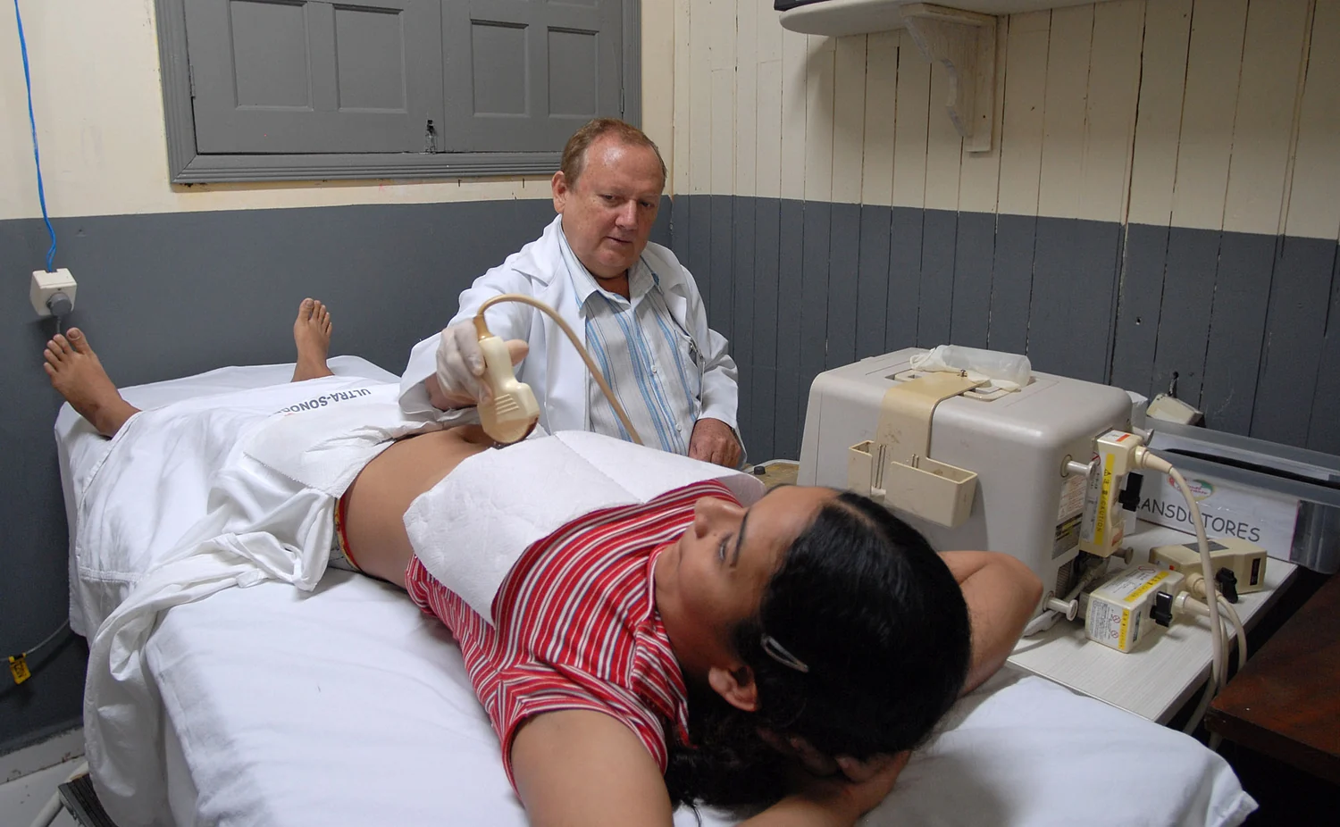
Why can a smooth-looking organ appear grainy on an ultrasound screen? The scanner is not simply photographing tissue: it sends sound into the body and listens for echoes. At any small region, many tiny structures can return waves that overlap. Some reinforce one another and some partly cancel. The resulting bright and dark flecks form speckle, a texture produced by wave behavior that can look like noise but also carries information about the tissue being imaged.
The scanner listens for echoes
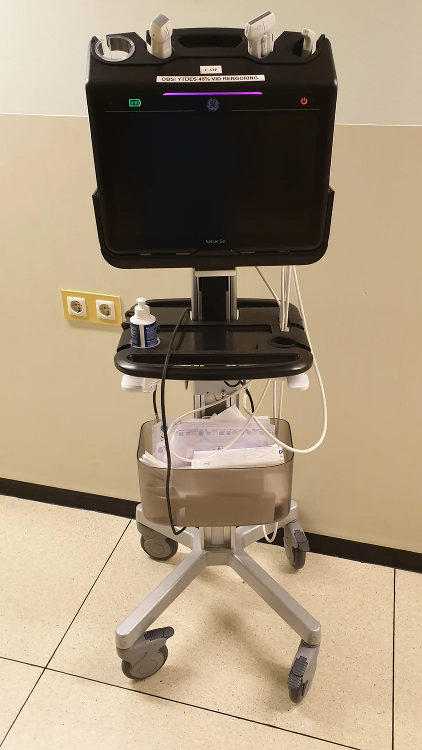
A transducer is the probe that both emits ultrasound and detects returning echoes. Its active elements commonly use piezoelectric materials: applying an electric field makes them vibrate and produce sound, while returning sound can produce an electrical signal. The scanner measures how long echoes take to return and uses the speed of sound to estimate how far away reflecting boundaries lie. Those distances help build a two-dimensional image. [1]
The probe needs good contact with the skin. Gel helps prevent small air pockets from forming between probe and skin, because air can block sound from passing into the body. Once sound enters, changes in acoustic properties at tissue boundaries send part of the wave back. A large, strong boundary can make a clear echo; many small features can produce a more complicated return.
When waves add up
Sound behaves as a wave, so echoes can combine. If two waves arrive in step, their pressure variations reinforce; if they arrive out of step, they partly cancel. Within a resolution cell—the small region from which the scanner combines echoes—many scatterers may contribute. Their different positions change the distances traveled and therefore the phases of the returning waves. The mixture of reinforcement and cancellation creates a mottled pattern of brighter and darker spots.
A useful mental experiment is to imagine several people tapping a table at slightly different times. Some taps coincide and sound louder; others fall between and soften the combined rhythm. Ultrasound echoes are not taps, and the scanner processes their timing, but the analogy captures why many individually tiny reflectors can yield a fluctuating combined signal. In tissue with many unresolved scatterers, the grain is a predictable consequence of summing waves, rather than a set of tiny objects the image can identify one by one.
Texture is signal and complication
Speckle can make boundaries harder to see. A faint structure may be obscured by nearby bright flecks, and the appearance can change with probe position or imaging settings. Yet speckle also reflects how sound interacts with the tissue inside a resolution cell. Image-processing methods may reduce its visual prominence, but smoothing can also soften fine detail. The challenge is not simply to erase grain; it is to preserve useful anatomy while making patterns easier to interpret.
The echoes themselves depend on the contrast between materials and on the geometry of their boundaries. Sound reflected from the interface between fluid and soft tissue, or between tissue and bone, contributes differently from sound scattered by small structures. Bone and air-containing tissues are difficult to image with standard ultrasound because sound does not pass through them as effectively as through many soft tissues. Thus a speckled image is shaped both by wave interference and by which parts of the body the beam can reach. [1]
What a model can clarify
Computational modeling means using mathematics, physics, and computer science to simulate a complex system. A model of ultrasound can represent sound propagation, tissue properties, scattering, and the way echoes are recorded. Researchers can alter one factor at a time in a simulation—for instance, scatterer distribution—while holding others steady. That can help explain why patterns change, and can suggest experiments for checking the explanation. [2]
But a simulated image is not a clinical measurement, and a convincing mechanism does not prove that a particular image feature diagnoses a condition. Models depend on assumptions and inputs; biological tissues contain variation that may be difficult to capture fully. In practice, ultrasound images are interpreted in context, and the technology is used for many purposes, from anatomical imaging to measurements of blood flow or tissue stiffness. Speckle is one clue about how the signal formed, not a verdict by itself. [1] [2]
Explore the physics: an explanatory diagram

Sources & further reading
- Ultrasound NIBIB
- Computational Modeling NIBIB
Prepared for the independent PSI Center reading library. About edition dates and source checking.
This explainer describes scientific principles. It does not assess individual treatment choices.