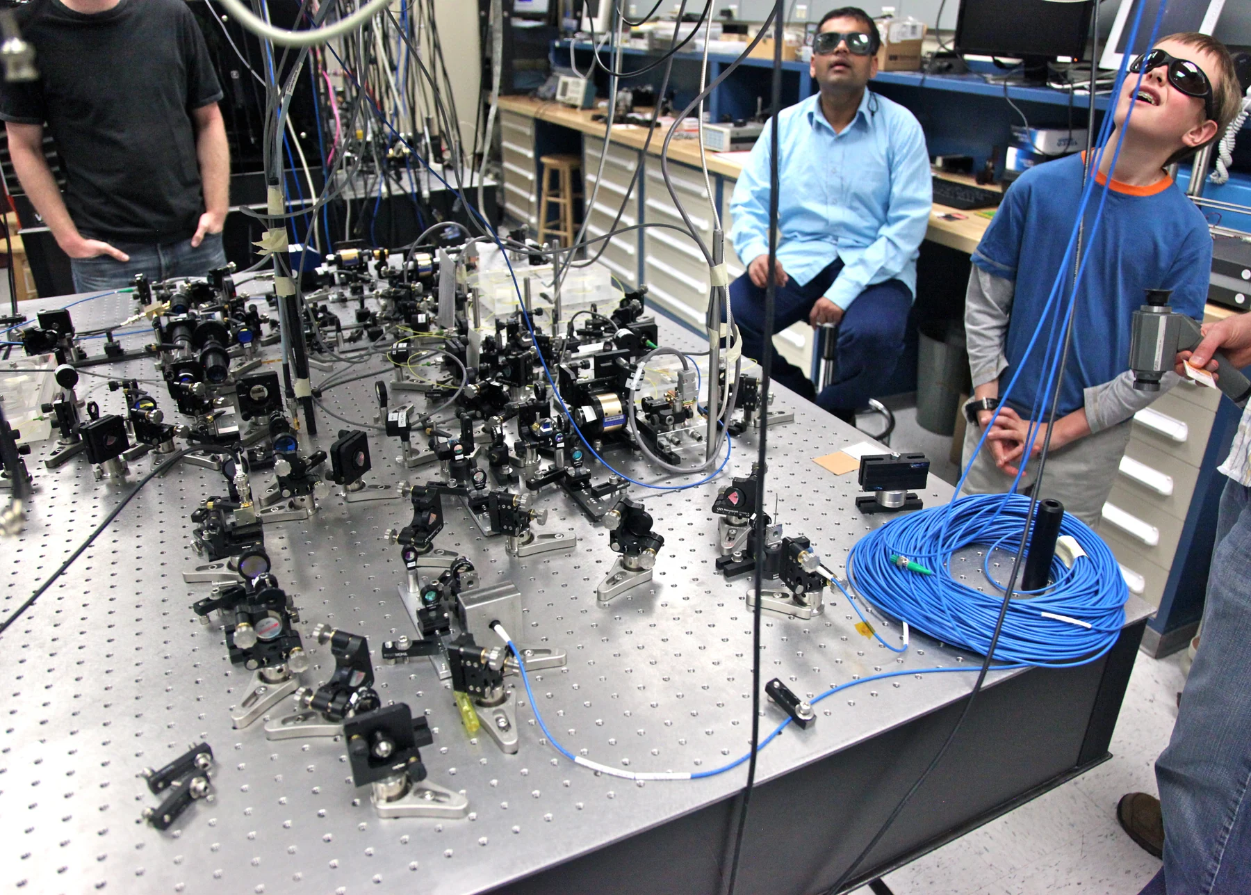
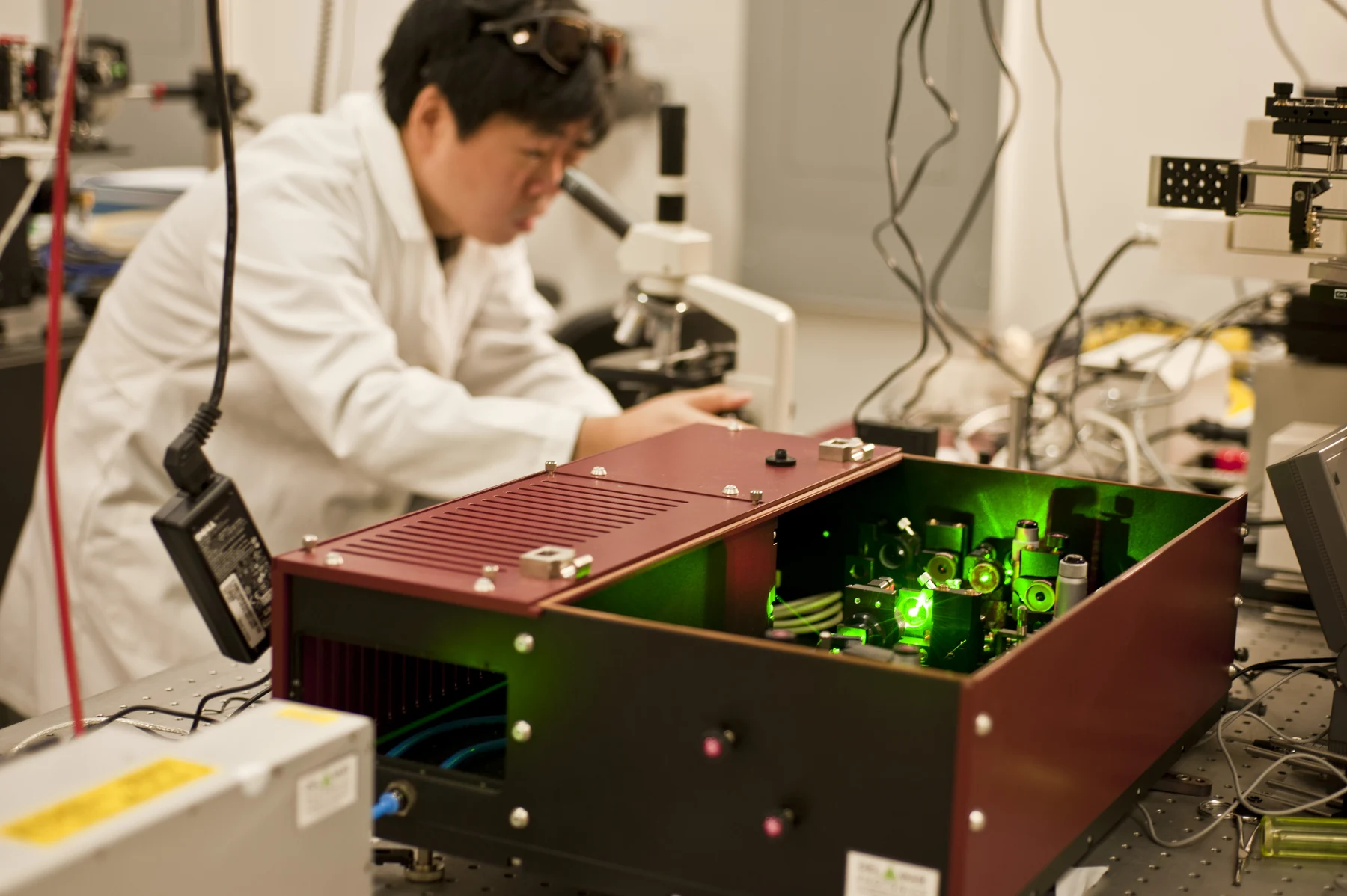
By tracking how light changes as it meets tissue, optical imaging can reveal structure and activity across scales. Its strength lies in choosing the right signal and understanding what that signal can—and cannot—show.
An imaging algorithm learns statistical patterns from examples. If those examples fail to reflect the people, scanners, or conditions encountered later, performance can become uneven, even when the training set is large.
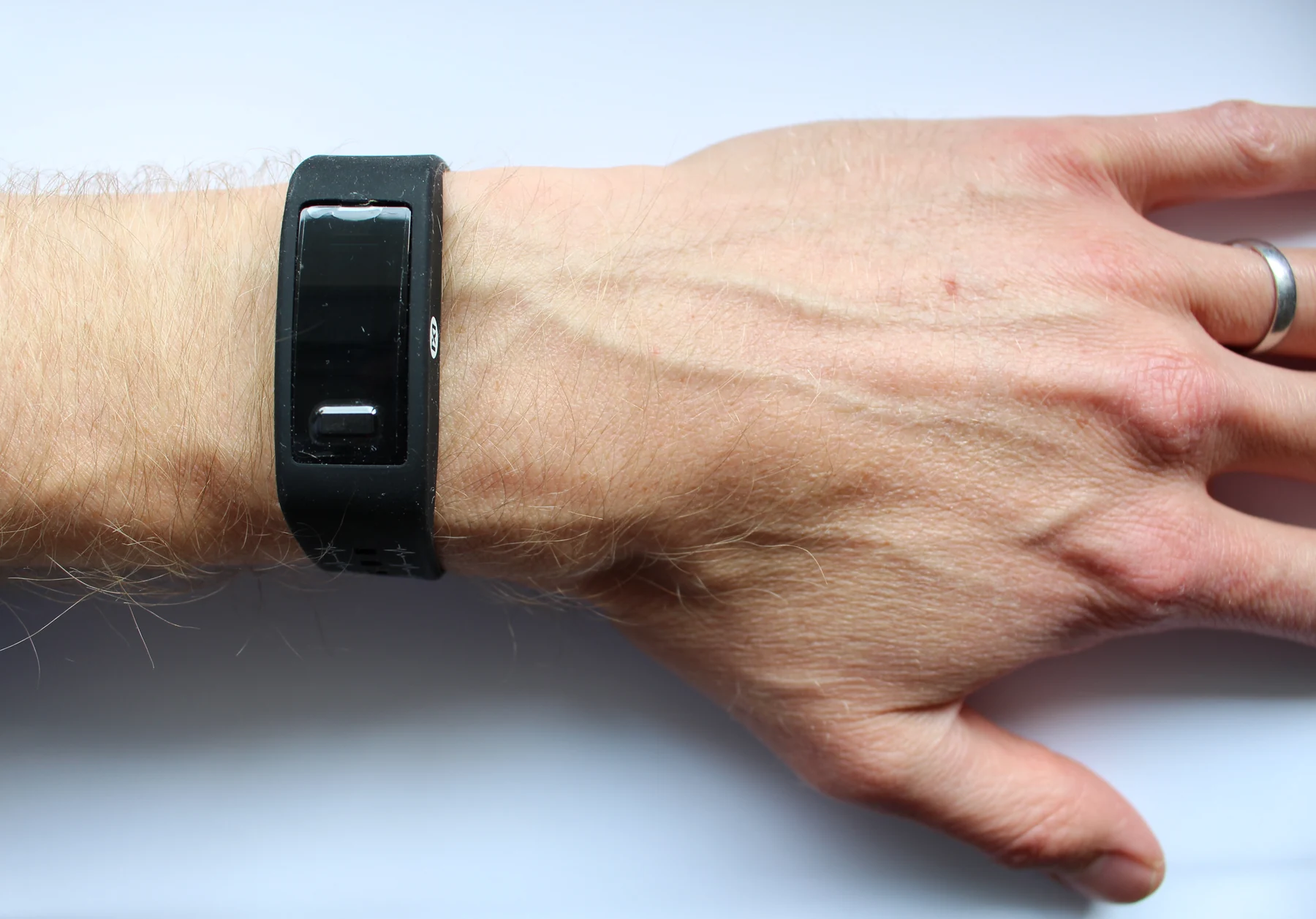
A wearable turns a bodily signal into data through sensors, software, and interpretation. Its value depends on whether those steps are accurate and whether using the information improves a meaningful health outcome.
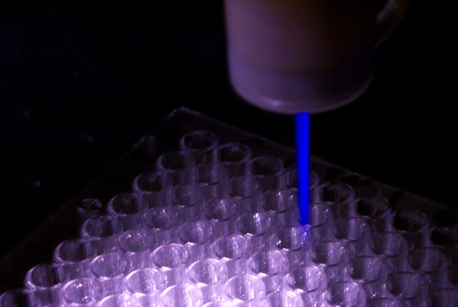
Photodynamic therapy relies on a paired sequence: a photosensitizing drug reaches target tissue, then selected light activates it. Wavelength, delivery route, oxygen chemistry, and limited light penetration shape its uses.
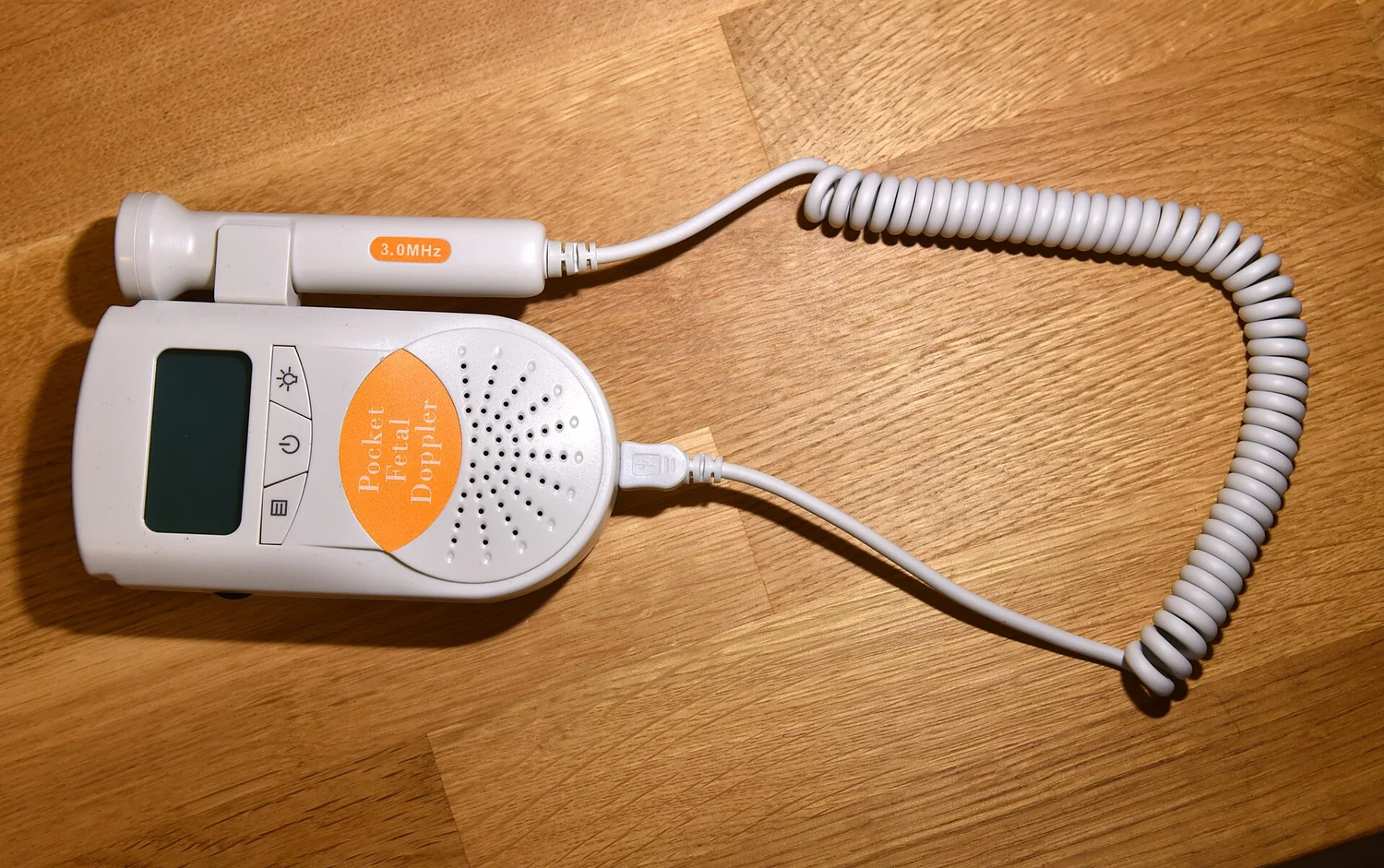
Frequency counts cycles per second; wavelength measures the distance a wave travels in one cycle. Their relationship helps explain ultrasound resolution and how electromagnetic radiation spans radio waves through x-rays.
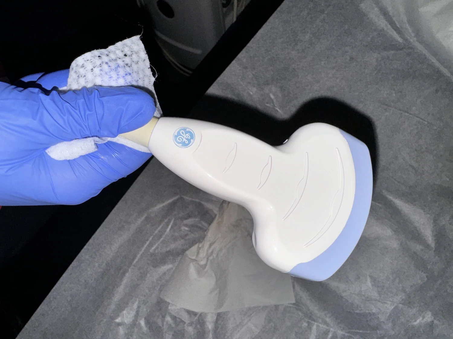
Ultrasound images form when sound echoes return from boundaries between tissues. Acoustic impedance helps explain why some interfaces reflect strong echoes, while sound also supports the very different process of hearing.
A plasma’s visible glow is only one part of what a device does. Gas composition, energy input, surroundings, and intended use determine whether the system sterilizes equipment or is being investigated for biological applications.
A wearable sensor measures signals through a changing, moving interface with skin and tissue. Motion can mimic or obscure physiology, so a reading needs context about both the body and device.
A plasma device’s gas is an active ingredient in the physics: it shapes which reactive particles form, how they move, and what they encounter. That makes treatment effects specific to the gas and setting.
PET maps where a radioactive tracer participates in bodily processes; CT supplies detailed structure. Their paired images help locate functional signals in anatomy, while each method contributes different information and limitations.
OCT uses reflected infrared light to build cross-sectional views of tissue beneath a surface. Its fine structural detail is valuable in the retina, while light scattering limits how deeply it can see.
A changing magnetic field can create a small electrical current in brain tissue without electrodes implanted there. The physics explains how rTMS reaches neurons, while clinical evidence and treatment use remain specific to particular conditions and protocols.
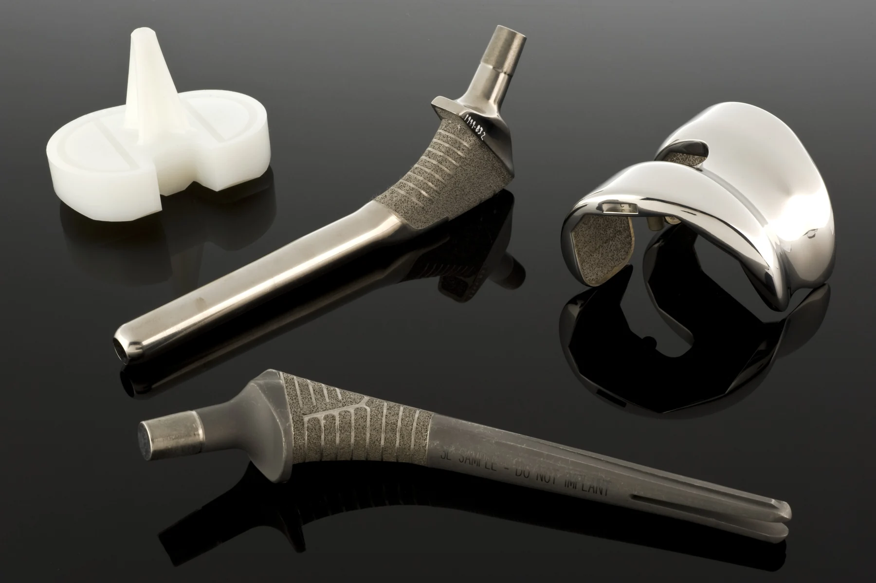
A prosthesis works through a moving boundary where materials, forces, and living tissue meet. Comfort and function depend on load transfer, fit, feedback, and the body’s response to an implanted or worn device.
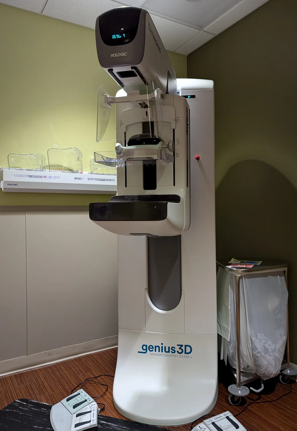
Contrast agents help imaging systems distinguish structures that would otherwise look similar. In CT and X-ray exams, their visibility comes from how they alter the passage of X-rays through selected parts of the body.
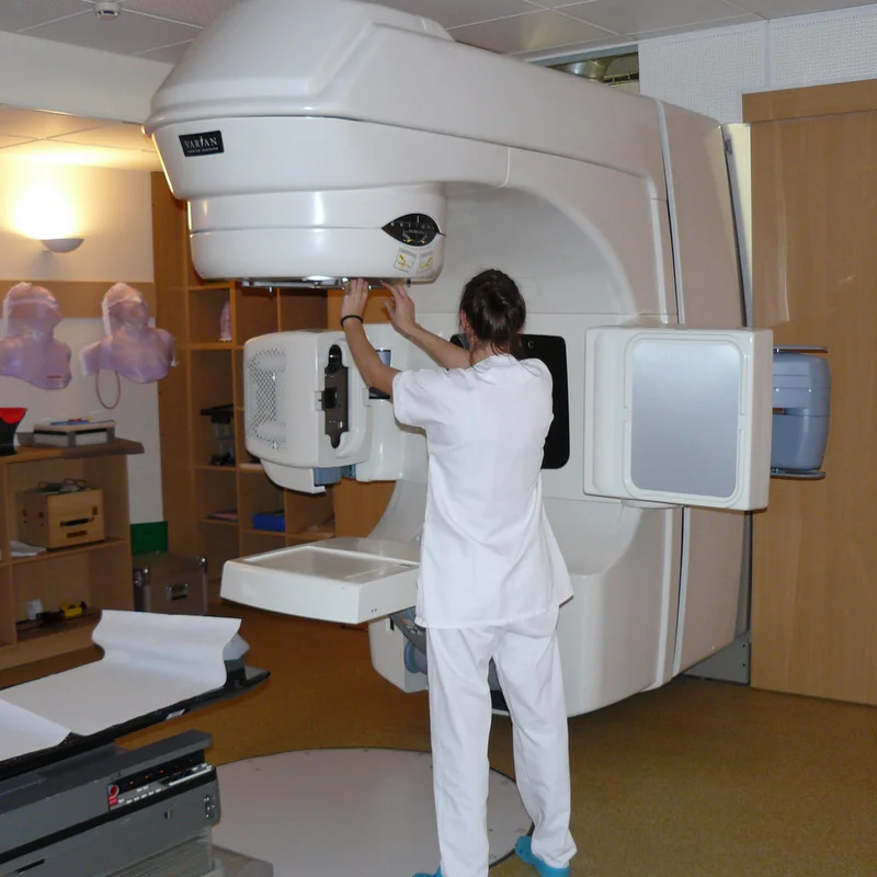
External beam treatment sends radiation from a machine into a planned region, while brachytherapy places a source inside the body. Understanding that physical difference explains why ordinary contact is safe after one treatment and precautions can matter with the other.
The knocks of an MRI scanner are a by-product of rapidly changing magnetic fields. Their force makes the machine vibrate, while the same fields help locate signals used to build detailed images.
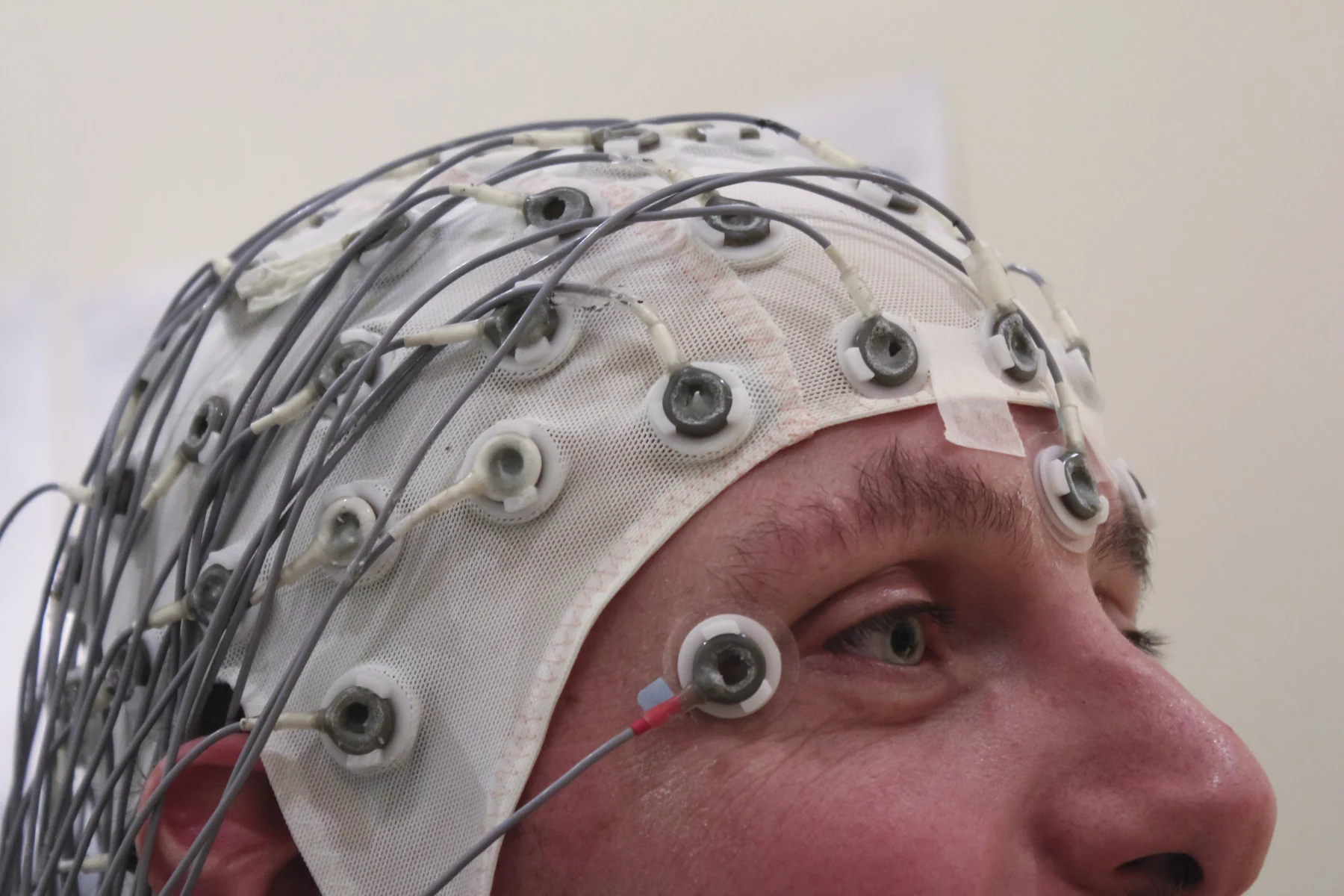
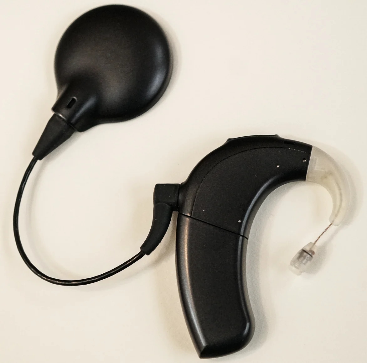
Sensors turn physical changes into readable signals, but every measurement captures only selected features of a system. Following sound from air vibration to brain activity shows how a signal can be transformed without being the whole experience.
A pacemaker senses electrical activity and delivers pulses when needed to support rate or timing. Its role makes sense only against the heart’s own electrical coordination, which organizes chamber contraction and helps blood move efficiently.