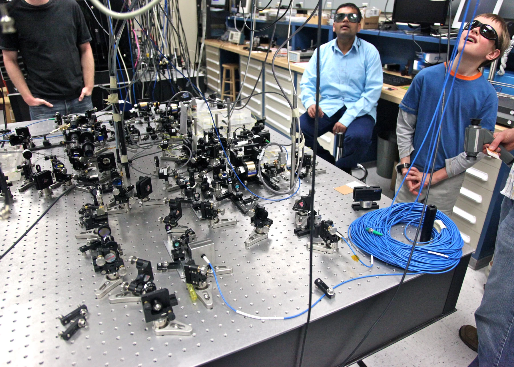
How can a beam of light reveal whether blood is flowing, tissue is changing, or cells are arranged in a particular way? The answer is that light does not simply pass through the body unchanged. Some is absorbed, some scattered or reflected, and some can be emitted after interacting with molecules. Optical imaging measures these altered signals and turns them into pictures or maps. What those maps mean depends on which light behavior a method is designed to detect. [1]
A signal is made by an interaction
Imagine shining a flashlight through a glass of clear water and then through a cloudy mixture. The first beam travels relatively directly; the second is scattered in many directions. Biological tissue behaves more like the cloudy mixture. Its molecules and structures absorb particular wavelengths, redirect light, reflect some of it, and in some cases emit light. Instruments collect selected parts of that response and use them to build an image. [1]
The distinction matters because an optical image is not a transparent photograph of everything inside the body. It is a measurement shaped by tissue composition, depth, wavelength, and the detector. A change in the signal may indicate a change in tissue properties, but interpreting it requires knowing what the method measures and how the signal travels. Optical techniques range from viewing tiny structures through a microscope to mapping properties of larger organs. [1]
Following a process means measuring a changing property
To follow a biological process, researchers can repeat measurements and compare the signal over time. For example, near-infrared light can be used in diffuse optical imaging to estimate properties such as total hemoglobin concentration and blood oxygen saturation in soft tissue. If the measured signal changes between observations, it can provide information about the tissue’s state. That information is a property map, rather than a direct movie of each blood cell moving. [1]
This is a useful distinction between observing a process and observing its consequences. A camera may record motion directly; an optical imaging system may infer that blood oxygenation or tissue structure has changed from the light returning to its detectors. The inference is valuable when the measured property answers the research or clinical question, but it does not automatically identify the cause of the change. [1]
Different optical methods answer different questions
Optical coherence tomography, or OCT, uses infrared light to produce cross-sectional images beneath a surface. Its comparison with ultrasound is helpful: both can form depth-resolved views from returned signals, but OCT uses light rather than sound. Ophthalmologists use it to image structures within the retina, while it can also help cardiologists assess coronary artery disease. The method is suited to detailed views of relatively shallow structures. [1]
Photoacoustic imaging begins with a different chain of events. A laser pulse is absorbed by tissue, that absorbed energy produces ultrasound waves, and a specialized detector records those waves. Because ultrasound can carry information from deeper tissue, this hybrid approach can help image blood vessels, tissue oxygenation, or blood-vessel growth in tumors. It combines optical contrast with acoustic detection; it is not simply a deeper version of OCT. [1]
Repeated views add context, not certainty
Light-based methods can be useful for repeated observations because visible, ultraviolet, and infrared light are less hazardous than some other forms of radiation, and optical techniques can measure several properties of soft tissue. Researchers may also combine optical imaging with methods such as MRI to obtain complementary information. Each method contributes a different kind of measurement, so agreement or disagreement between them can sharpen the questions scientists ask. [1]
The idea of following a process also appears in rapid diagnostics, where a biological sample is converted into a result quickly, often near the point of care. These tests illustrate a related measurement principle: a signal becomes useful through a defined relationship to the question being asked. A rapid test is not the same as imaging tissue, but both require an interpretable link between what an instrument detects and the biological feature of interest. [2]
A good optical image therefore starts with a precise question: which tissue property might change, at what depth, and on what scale? A microscope can resolve cellular detail; a diffuse optical method can estimate oxygenation across soft tissue; OCT can show layered structure. Matching the instrument to the question makes it possible to track change while keeping the interpretation tied to what the measurement actually records. [1]
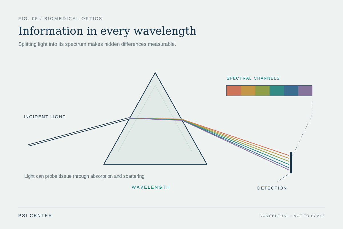
Explore the physics: an explanatory diagram
Sources & further reading
- Optical Imaging NIBIB
- Rapid Diagnostics NIBIB
Prepared for the independent PSI Center reading library. About edition dates and source checking.
This explainer describes scientific principles. It does not assess individual treatment choices.


