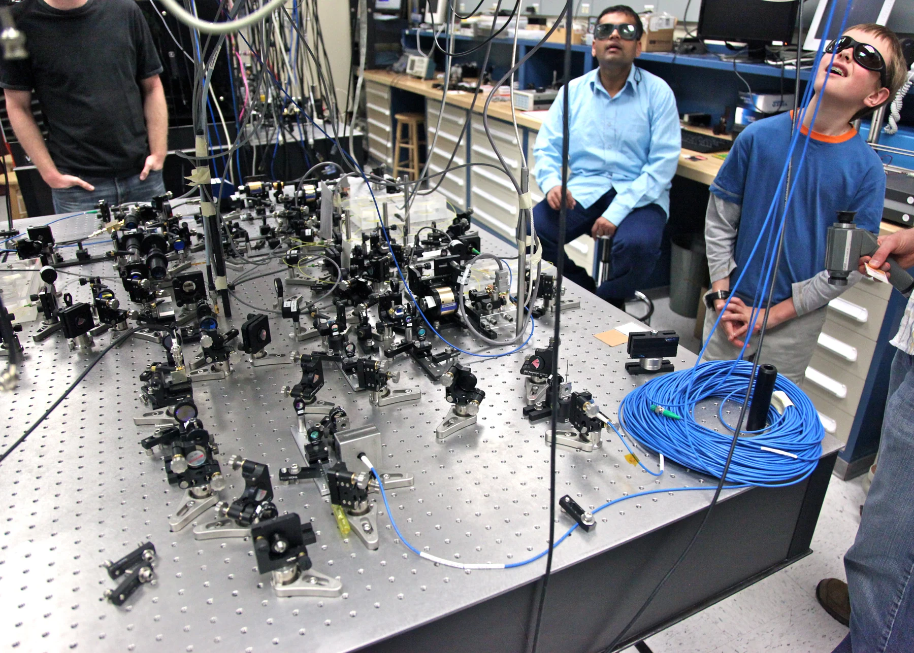
What could a clinician learn by pairing light with a magnetic field? Optical imaging detects how light is absorbed, scattered, reflected, or emitted by tissue, while MRI reads signals from protons responding to magnetic fields and radio waves. These are distinct physical interactions. A combined approach can place different kinds of information side by side, offering a richer view than either signal may provide alone.
Light carries clues from tissue
When light enters biological material, it does not simply travel straight through. Molecules absorb some wavelengths, structures scatter light in different directions, and some substances emit light. Optical imaging uses these interactions to visualize biological samples from molecules and cells to tissues and organs [1]. Different techniques work at different scales: microscopy magnifies tiny structures, while optical methods such as diffuse optical imaging can measure tissue properties including hemoglobin concentration and blood oxygen saturation [1].
A mental picture is a flashlight shining into fog. The returned light depends on what is in the fog and how it is arranged, but scattering blurs the path. In tissue, that behavior can provide useful information about soft tissue, though it also influences how deeply and clearly light can probe. Optical coherence tomography uses infrared light to make subsurface cross-sectional images, while photoacoustic imaging uses laser pulses that generate ultrasound waves in tissue [1].
MRI listens to proton behavior
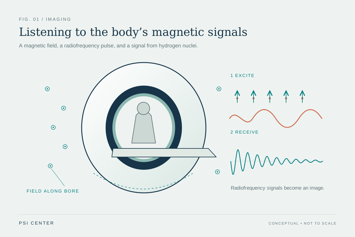
MRI uses a strong magnet to align protons, especially those in water molecules, within the body. A radiofrequency pulse disturbs that alignment; when the pulse stops, sensors detect energy released as protons return toward the magnetic field’s direction. The timing and amount of the signal vary with the surrounding molecular environment, allowing different tissues to be distinguished [2]. MRI does not use ionizing x-rays, and it is particularly useful for imaging soft tissues such as the brain, muscles, ligaments, and tendons [2].
MRI and optical imaging therefore answer different physical questions. Optical methods track how tissue interacts with light. MRI measures signals associated with proton alignment and relaxation. Neither method is a universal window into the body: the image reflects the properties each modality can detect, and the body’s structure determines how those signals are produced.
Why combine unlike signals?
A map from one modality can leave uncertainty that another modality helps address. Optical imaging is particularly useful for measuring multiple properties of soft tissue, and it can be combined with MRI to provide enhanced information for monitoring complex diseases or for research [1]. For example, optical measures of oxygenation could complement MRI’s detailed anatomical image: the anatomy gives a view of structure, while the optical signal can add information about a tissue property.
This does not mean the methods merge into one signal or automatically improve every examination. A combination is useful only when the added information speaks to the question being asked. One can imagine overlaying two transparent maps: one outlines streets, the other marks elevation. Their value comes from complementarity, but each map still has its own resolution and limitations.
Different physics bring different constraints
Optical methods can be repeated because visible, ultraviolet, and infrared light are less hazardous than ionizing radiation, according to NIBIB [1]. MRI also avoids ionizing radiation, but it uses a powerful magnetic field that can exert strong forces on certain metal objects. Patients need to disclose implants before an MRI scan, and the scanner’s loud sounds may require ear protection [2]. The safety considerations arise from the physical energy and equipment involved, not from the label “imaging” alone.
For a combined system, practical design must account for both modalities’ requirements: how to collect light signals and how to operate within or alongside an MRI environment. The basic idea is simple—different physical probes can reveal different tissue properties—but a useful clinical image still depends on careful measurement, interpretation, and the specific task. MRI is not plasma or x-ray imaging, and optical methods do not by themselves provide every kind of anatomical detail.
Explore the physics: an explanatory diagram
Sources & further reading
- Optical Imaging NIBIB
- Magnetic Resonance Imaging NIBIB
Prepared for the independent PSI Center reading library. About edition dates and source checking.
This explainer describes scientific principles. It does not assess individual treatment choices.