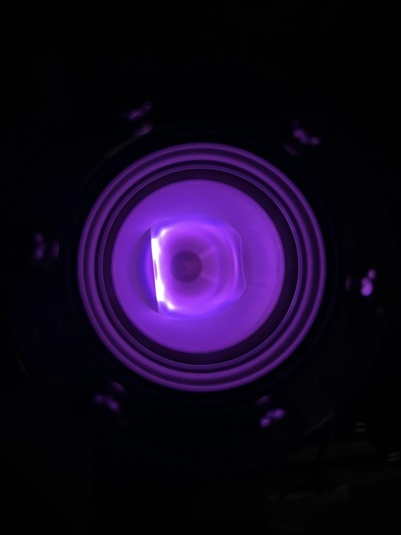
Cold Plasma: Chemistry, Dose, and Wound Healing
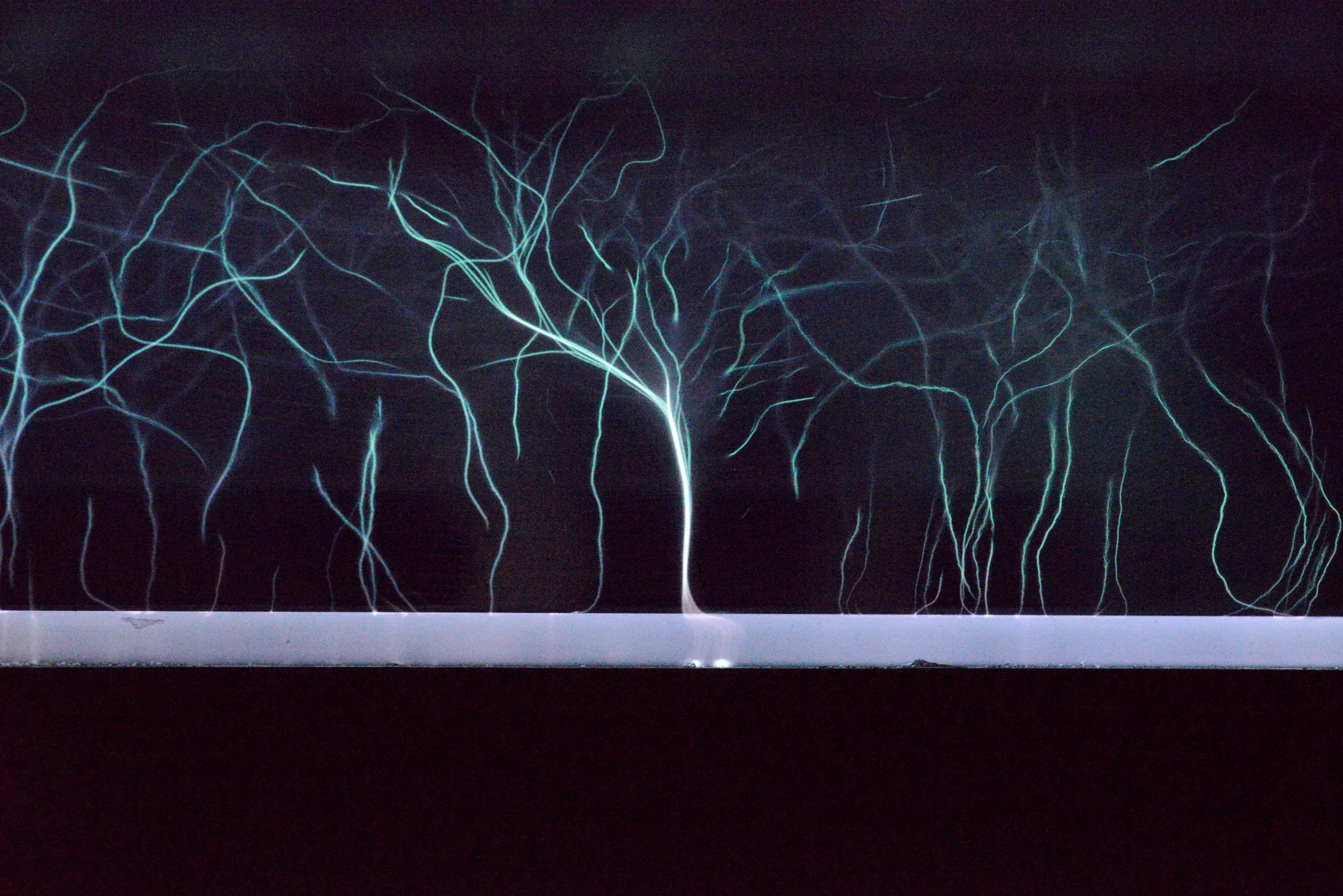
A close examination of how a cool electrical discharge could influence wound repair, what randomized trials actually measured, and why seconds of exposure do not define a transferable biological dose.
A closer look at the science
behind better healthcare.
Follow a question from first principles to the clinic. Browse the science of imaging, treatment, materials, and measurement.
324 articles
Deep dives first, then reading editionsA close examination of how a cool electrical discharge could influence wound repair, what randomized trials actually measured, and why seconds of exposure do not define a transferable biological dose.
Follow the signal from a small imbalance in nuclear spins to spatial encoding and reconstruction, with worked examples showing why frequency, resolution, field of view, noise, and scan time are different quantities.
A better dose distribution is a powerful starting point. Range uncertainty, tissue biology, and contrasting randomized trials explain why it is only the beginning of the clinical argument.
How focused sound deposits heat or drives mechanical bubble activity, why imaging and motion control matter, and how indication-specific FDA authorizations differ from evidence of clinical benefit.
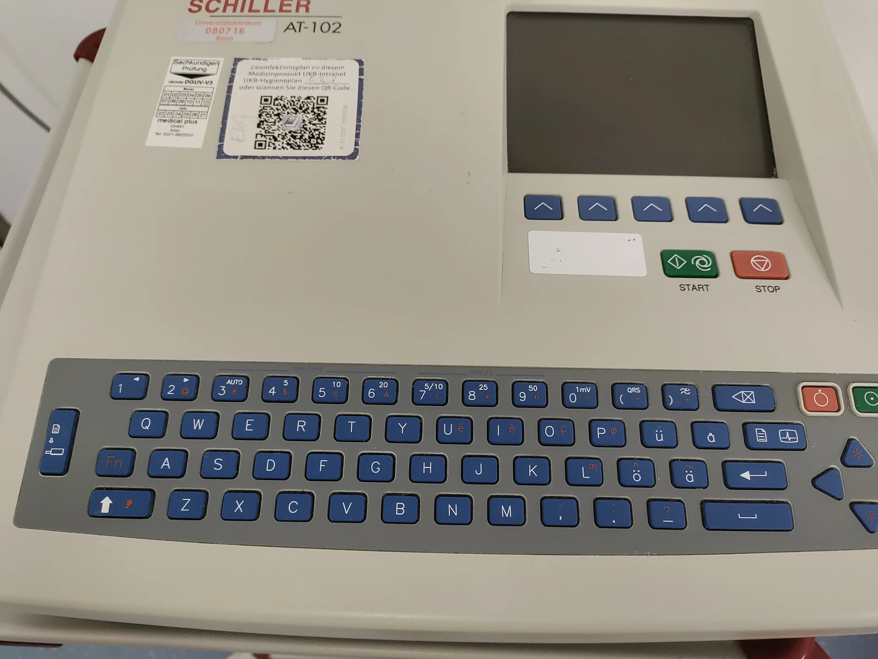
Cells use charged particles to coordinate activity, and the heart turns that coordination into a repeating pump cycle. An electrocardiogram reads the resulting pattern from the skin without sending current into the body.
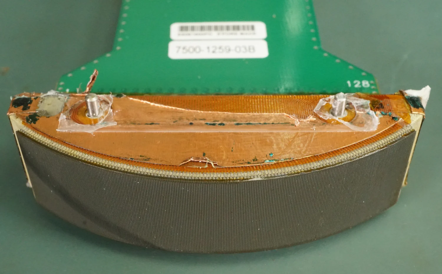
Ultrasound is sound above human hearing, but its medical uses depend on how waves travel, reflect, and deliver energy. Imaging and therapy use different intensities and goals.
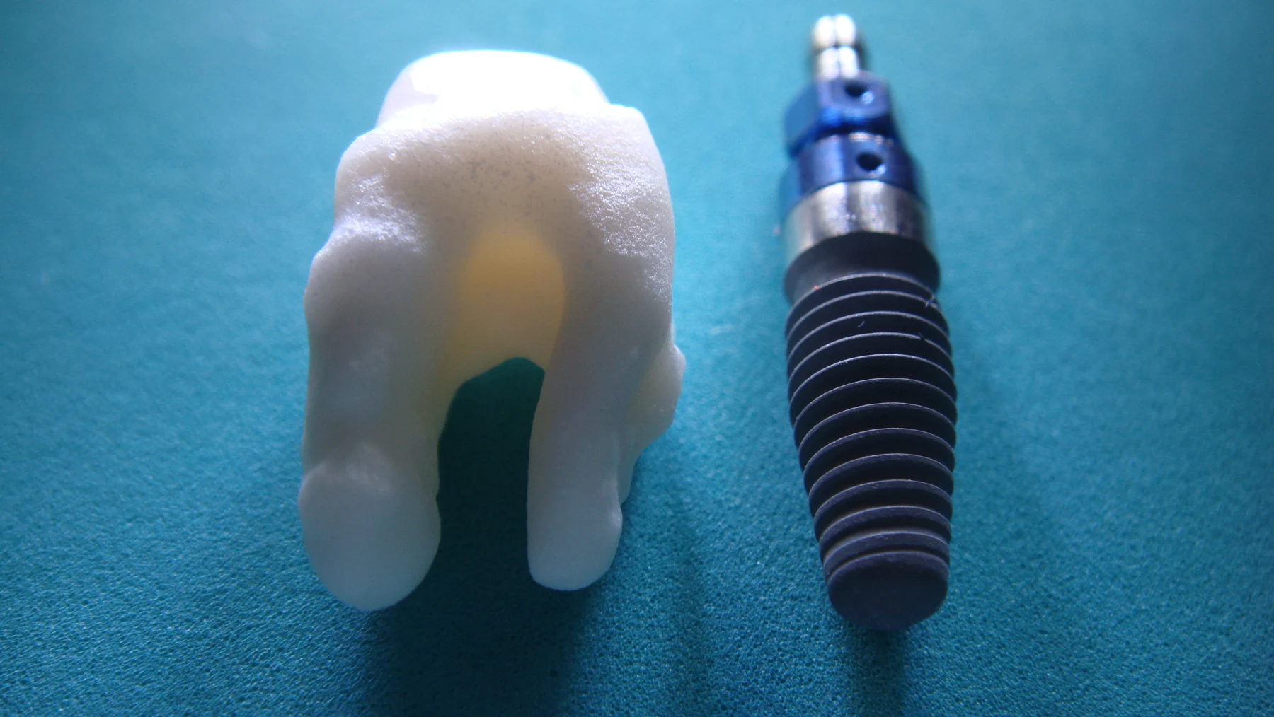
A biomaterial earns its name through its intended interaction with the body, not simply through its chemical category. Its performance depends on placement, function, manufacturing, and the body’s response over time.
A plasma begins when some gas electrons become free, making the mixture responsive to electric and magnetic fields. That shared property links lightning, stars, and carefully controlled sterilization equipment.
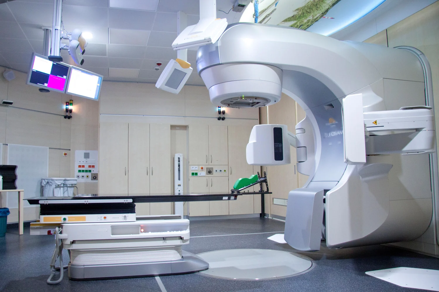
Ionizing radiation carries enough energy to disrupt atoms and damage DNA. In cancer therapy, that damage can stop cell division over time, while injury to nearby healthy cells explains side effects.
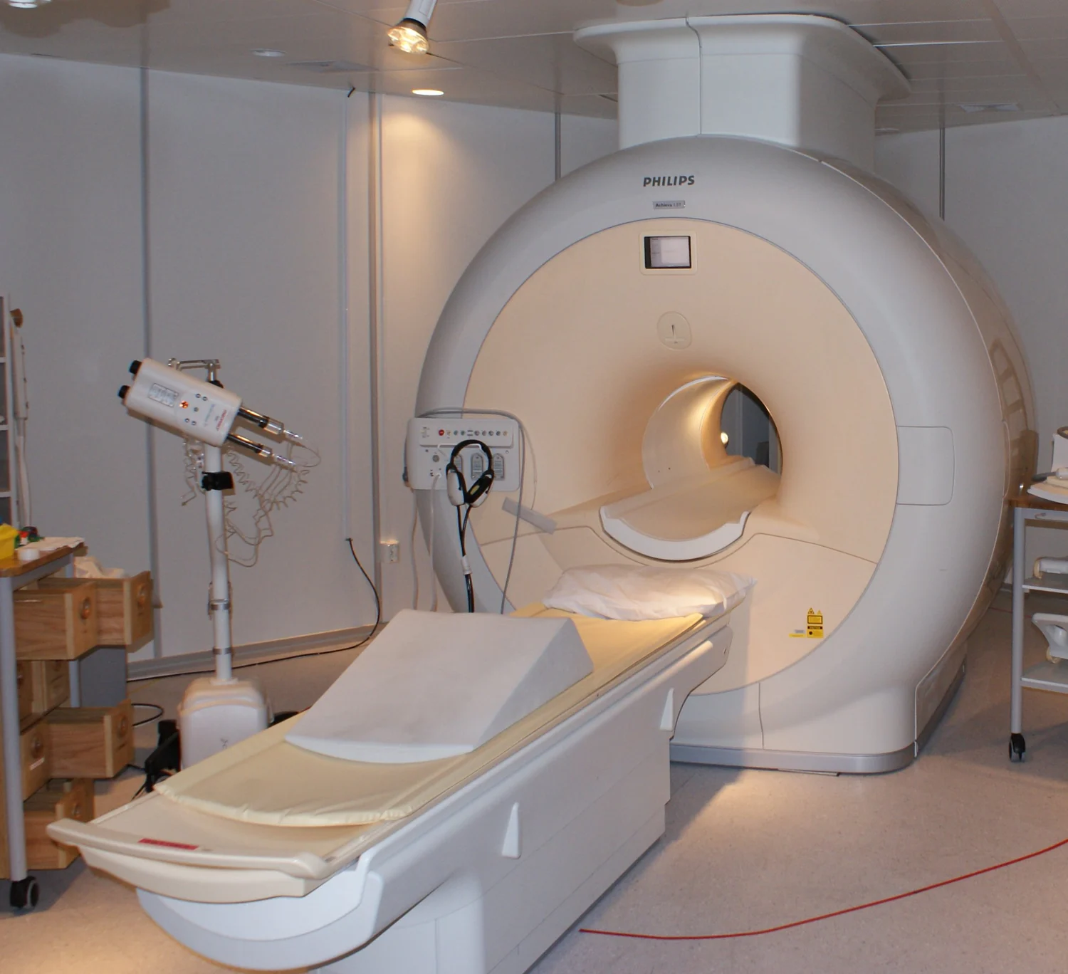
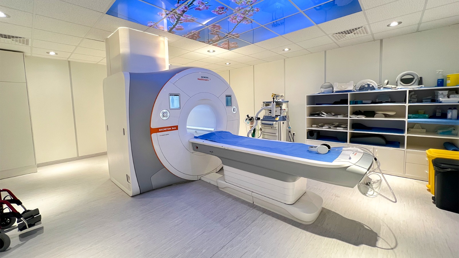
MRI converts tiny changes in hydrogen-proton behavior into spatial detail. Magnetic fields align the signal, radiofrequency pulses disturb it, and carefully timed measurements distinguish tissue environments without x-rays.
Light can scatter, reflect, absorb, or emerge from tissue, and each interaction carries different information. Wavelength, focus, and geometry shape both optical images and laser effects.
Energy measures a total transfer, while power describes how quickly that transfer occurs. In treatment and imaging, timing and location help determine how energy interacts with tissue, so a single number rarely tells the whole story.
MRI contrast comes from how protons respond and recover in different tissue environments, as well as from scan settings and image processing. Optical imaging measures light interactions and can add complementary information at other scales.
An implanted electronic device must sense and deliver carefully timed signals while sharing space with external magnetic fields. Compatibility is about keeping those intended functions reliable in the environments people encounter.
Radioactivity counts how often unstable atoms transform; radiation dose describes energy deposited in matter and its potential biological significance. Keeping those measures separate helps make sense of medical imaging, radioactive materials, and risk.
Digital anatomy can guide a device whose shape fits an individual body, but a close geometric match answers only one design question. Materials, manufacturing, function, and evidence still matter.
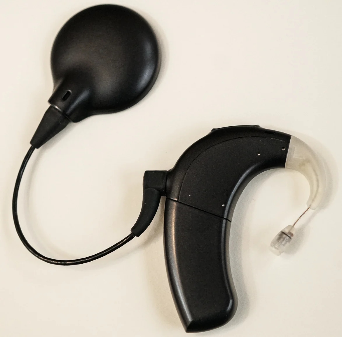
Hearing depends on a chain of mechanical and neural steps, so making sound louder cannot solve every hearing problem. Comparing hearing aids with cochlear implants shows where that chain can break.
IMRT varies beam strength across many small regions and directs beams from several angles. Planning software uses images to shape the dose distribution, while healthy tissue along each beam path still matters.