How does a heart muscle know when to squeeze, and how can a sensor on the skin detect that instruction? The answer begins with charged particles moving across cell membranes. In the heart, those tiny electrical changes travel through tissue in an ordered sequence, coordinating chambers so blood moves forward. The body does not generate electricity as a separate substance; its electrical signals are changes in voltage created by the movement of ions in and around living cells.
A voltage begins at a cell membrane
A cell’s membrane separates its interior from the fluid around it. Charged atoms and molecules, called ions, are distributed unevenly on the two sides. Because opposite charges attract, this separation creates an electrical voltage across the membrane, much like a very small, flexible battery. Channels in the membrane open or close to let selected ions pass. When that happens, the local voltage changes.
In excitable cells such as nerve and heart muscle cells, a voltage change can trigger a wave of further changes in neighboring membrane. This traveling event is called an action potential. It is not a stream of electricity flowing down a wire: it is a coordinated sequence of ion movements that renews itself along the cell. The resulting signal can prompt a muscle cell to contract.
The heart uses signals to time its pump
The heart’s electrical system controls the rate and rhythm of each beat. Its signals coordinate contractions so the chambers squeeze in a useful order and blood continues through the circulation [1]. Imagine a line of people passing buckets: if everyone moves at random, little reaches the end; if each person acts in sequence, the transfer works. The heart’s electrical timing serves a similar coordinating role, though its real mechanism is cellular and far faster.
The timing matters because contraction is only one part of pumping. The chambers must also relax and fill. Electrical activity is therefore linked to a mechanical cycle: a change in voltage spreads through heart tissue, muscle contracts, and then the tissue recovers so the next cycle can begin. Disturbances in the heart’s electrical system can make pumping less effective even when the muscle itself is not the only issue.
Skin electrodes listen to the heart
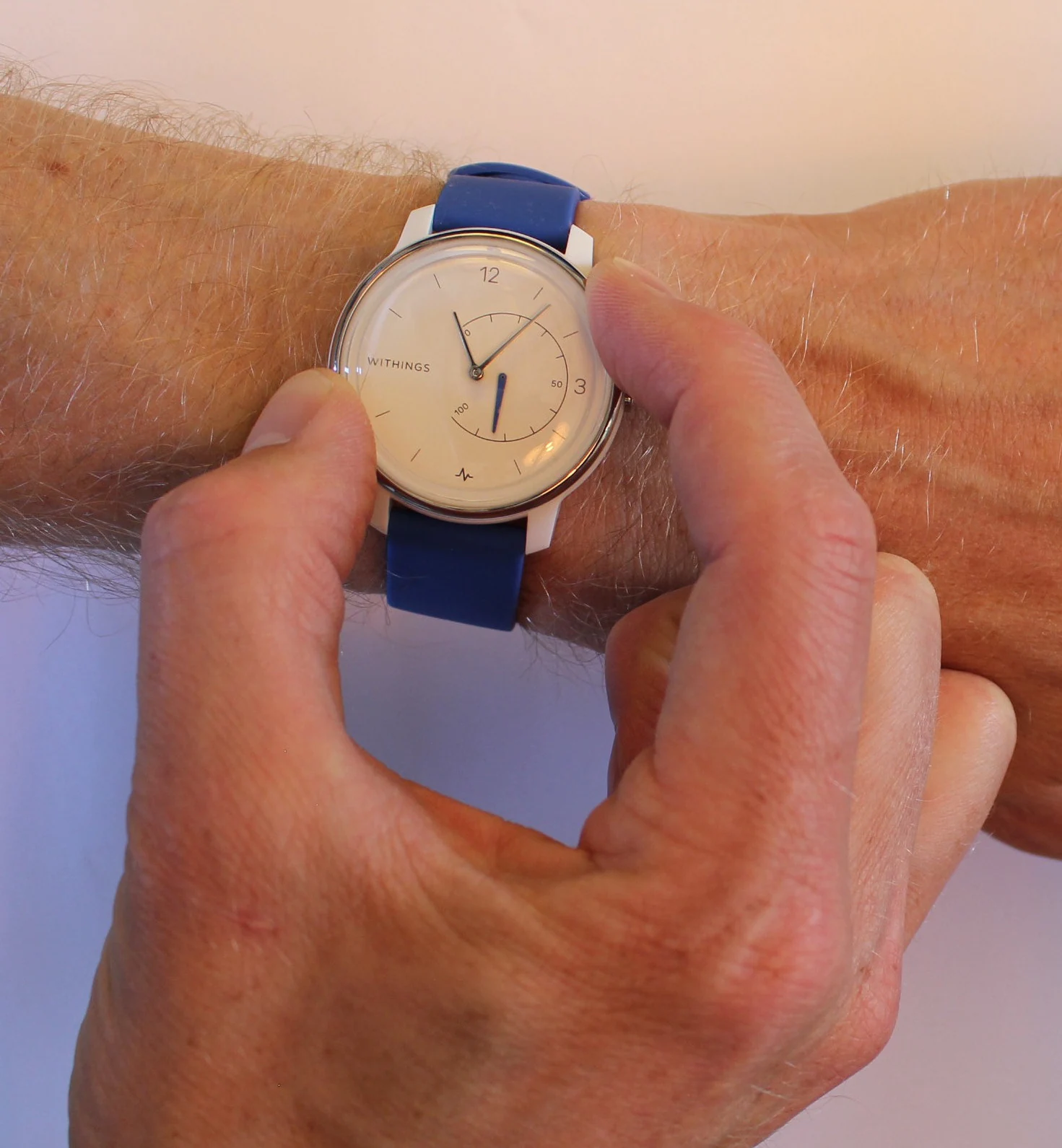
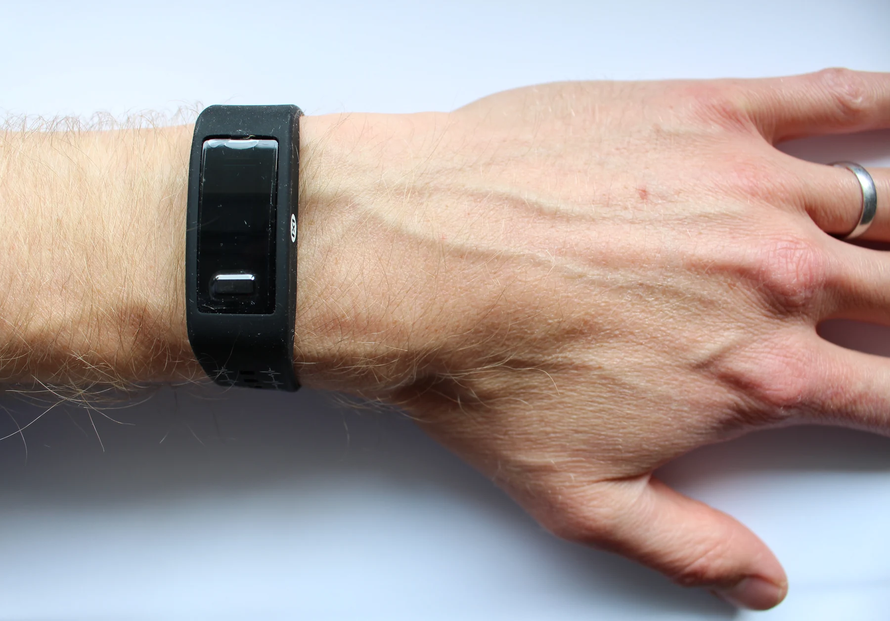
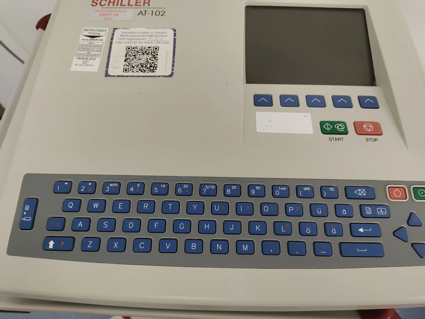
An electrocardiogram, also called an ECG or EKG, records electrical signals from the heart using small sensors attached to the skin. It does not send electricity into the body. Instead, electrodes detect tiny voltage differences produced as the heart’s electrical activity spreads through tissue and body fluids [2]. The machine displays those changing differences as lines on a screen or paper.
A familiar ECG trace has a P wave, a QRS complex, and a T wave. These portions correspond to different stages of the heart’s electrical cycle, including activation of the upper chambers, activation of the lower chambers, and recovery after contraction. The trace is a measured pattern, not a direct picture of a single cell or a forceful squeeze. Its shape depends on how electrical activity reaches the electrodes.
A useful signal has limits
An ECG can show heart rate, rhythm, and the timing and strength of electrical signals as they pass through different parts of the heart. Clinicians interpret the pattern alongside symptoms, medical history, and other information; an ECG result alone does not diagnose every heart condition [2]. A short recording is also a snapshot. If an unusual rhythm comes and goes, it may not occur during those few minutes, so a longer recording may be used.
The same principle reaches beyond the heart: living tissues rely on electrical differences to communicate and respond. Sensors can measure some of those differences, but a reading always reflects a particular place, time, and measurement setup. Understanding that distinction helps explain both the power and the limits of bioelectric measurements: they reveal an organized physical process without turning every line on a display into a complete account of health.
Sources & further reading
- How the Heart Works NHLBI
- Electrocardiogram MedlinePlus
Prepared for the independent PSI Center reading library. About edition dates and source checking.
This explainer describes scientific principles. It does not assess individual treatment choices.