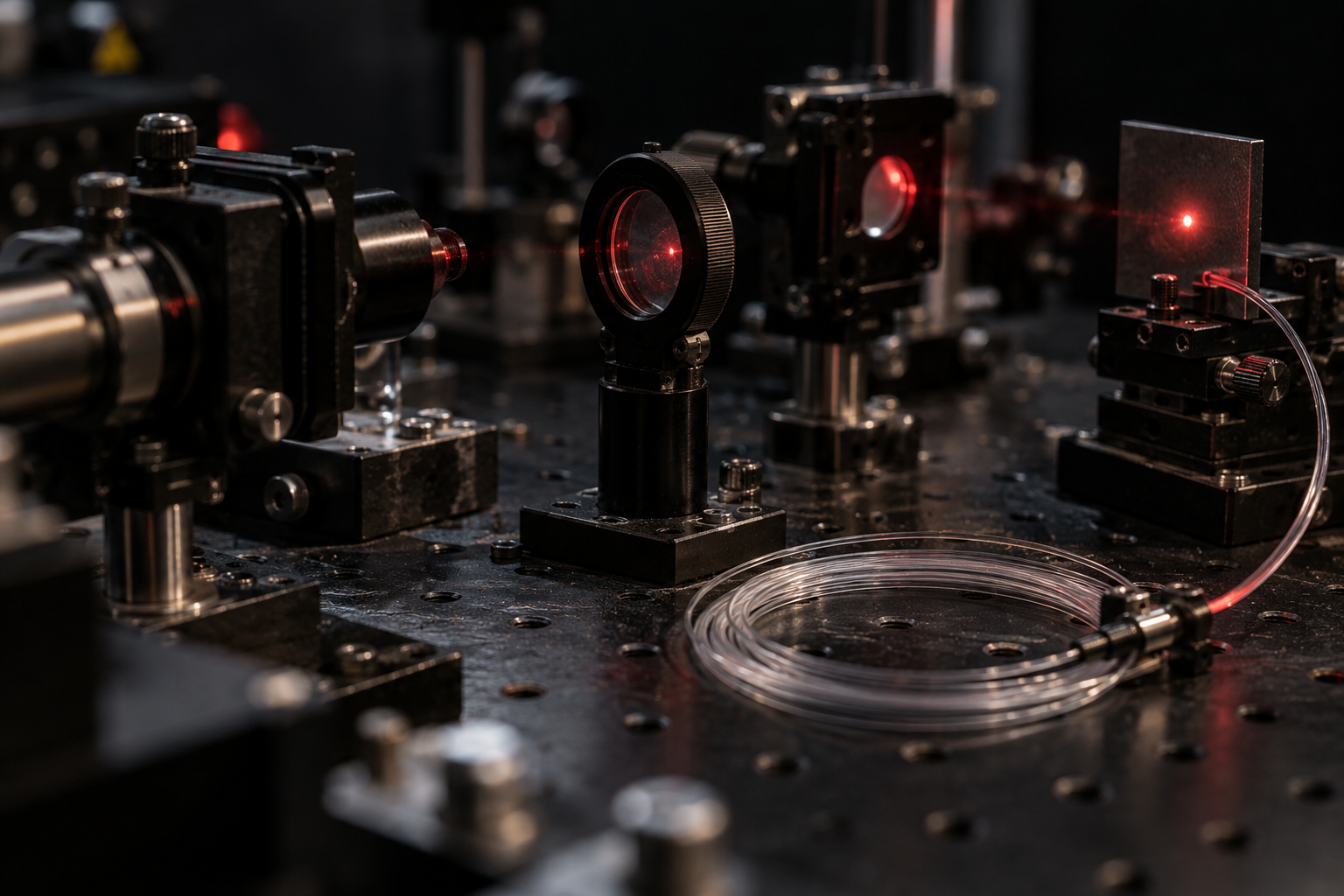
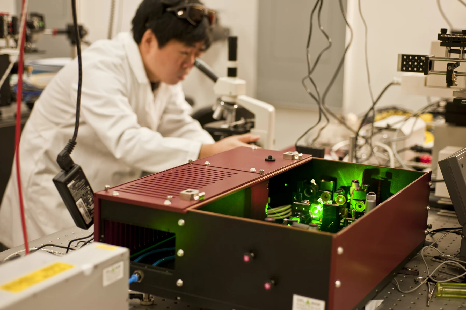
What happens when a beam of light enters living tissue? Some photons are absorbed, some change direction, and some return toward a detector. Those interactions determine whether light reveals a surface, probes deeper structure, or delivers concentrated energy. Optical imaging turns these varied responses into information about cells and organs. A laser adds a distinctive feature: its light can be concentrated into a narrow, directional beam, making control of where energy lands especially important.
A photon’s possible paths
Light interacting with tissue can be absorbed, scattered, reflected, or emitted. Absorption transfers energy to molecules; scattering redirects light; reflection sends a portion back at a boundary; emission occurs when matter releases light after excitation. Optical imaging uses these behaviors to visualize biological material at scales ranging from molecules and cells to whole tissues [1].
Imagine shining a flashlight through a fogged glass. The beam may be redirected by particles, partly absorbed, and partly transmitted. Tissue is much more complex than glass, but the comparison helps explain why a detector receives a mixture of signals rather than a simple shadow. Different tissues absorb and scatter light differently, which can provide clues about their composition and function [1].
Wavelength and depth shape the view
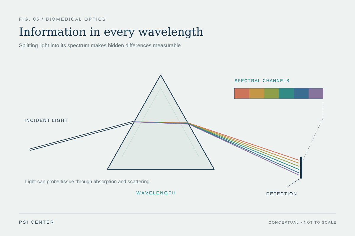
Visible, ultraviolet, and infrared light are all electromagnetic radiation, but they differ in wavelength and energy. The choice affects what tissue features can be measured and how far light can travel before scattering or absorption overwhelms useful detail. Optical coherence tomography uses infrared light to create cross-sectional views beneath a surface, including detailed images of the retina [1].
Diffuse optical methods use near-infrared light to estimate properties such as hemoglobin concentration and blood oxygen saturation in soft tissue [1]. These measurements are not a literal photograph of every cell: they infer tissue properties from how light has traveled and changed. Optical techniques can be useful for repeated monitoring because these forms of light are less hazardous than ionizing radiation [1].
When light makes sound
Photoacoustic imaging combines optical excitation with ultrasound detection. A brief laser pulse is absorbed in tissue; the resulting local energy deposition produces a small pressure wave that travels as ultrasound and can be measured by a transducer [1]. Thus, light supplies contrast while sound helps carry information from deeper tissue, where detecting photons directly can be difficult.
The method illustrates how one kind of wave can be converted into another. Blood absorbs light differently from surrounding structures, so photoacoustic signals can map vessels and oxygenation-related properties. NIBIB describes research using this approach to image blood flow in hands and feet, with further applications still being explored [1]. A promising measurement capability is not by itself proof of improved clinical outcomes.
A focused beam is a different tool
Laser light has a specific wavelength and can be amplified into a narrow beam traveling mainly in one direction. Concentrating light into a small area can create high intensity even at a distance [2]. This differs from an ordinary lamp, which emits a mixture of wavelengths in many directions. Lasers therefore serve roles from measurement and communication to medical procedures.
The same concentration that makes a laser useful also means exposure risks depend on the product and its use. FDA classifies laser products by hazard; higher classes can pose greater risks to eyes or skin, and some powerful products can present fire hazards [2]. Lasers designed for medical procedures must also meet medical-device requirements [2]. In tissue, a beam’s effects depend on wavelength, intensity, focus, exposure, and the properties of the target—not on the word “laser” alone.
Explore the physics: an explanatory diagram
Sources & further reading
Prepared for the independent PSI Center reading library. About edition dates and source checking.
This explainer describes scientific principles. It does not assess individual treatment choices.