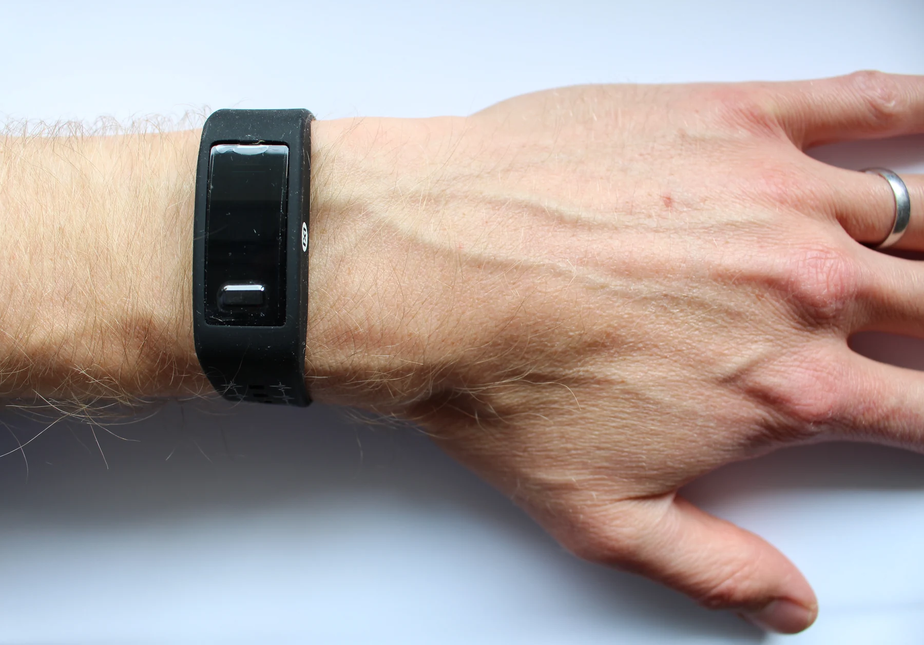
Pulse oximeters infer oxygen saturation from light, but skin pigment and other conditions can affect the signal. Understanding the measurement helps explain both accuracy gaps and how researchers are trying to improve them.
Microfluidic diagnostics guide tiny fluid volumes through channels where biological chemistry can become a readable signal. Their compactness is useful, but the result still depends on what the test detects and how reliably it distinguishes conditions.
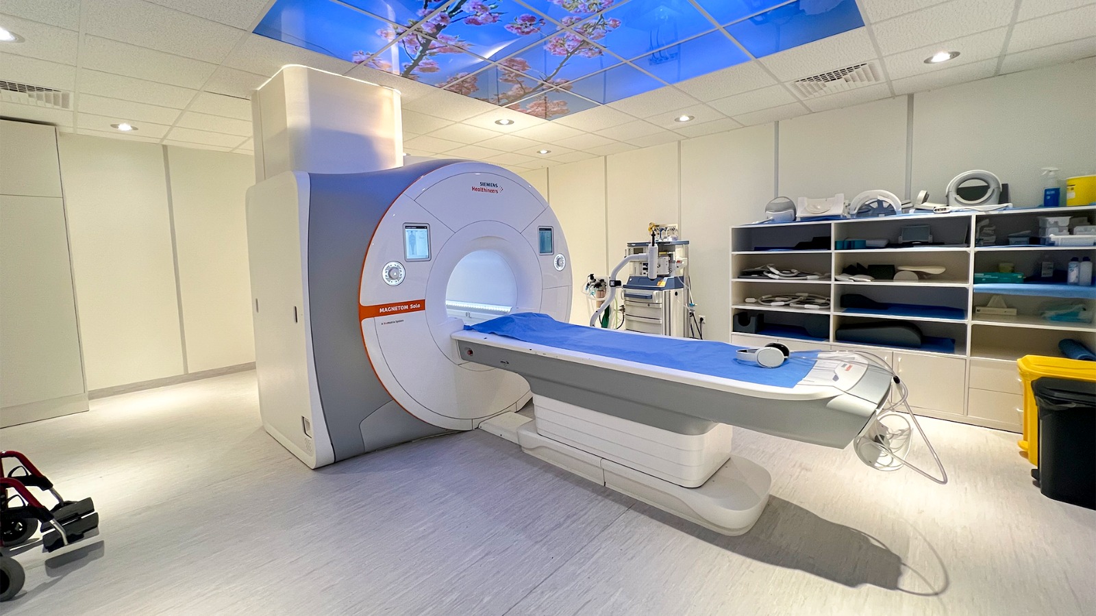
MRI maps tissue through magnetic behavior, while optical imaging measures how light is absorbed, scattered, reflected, or emitted. Together, these methods can reveal different aspects of the same biological problem.
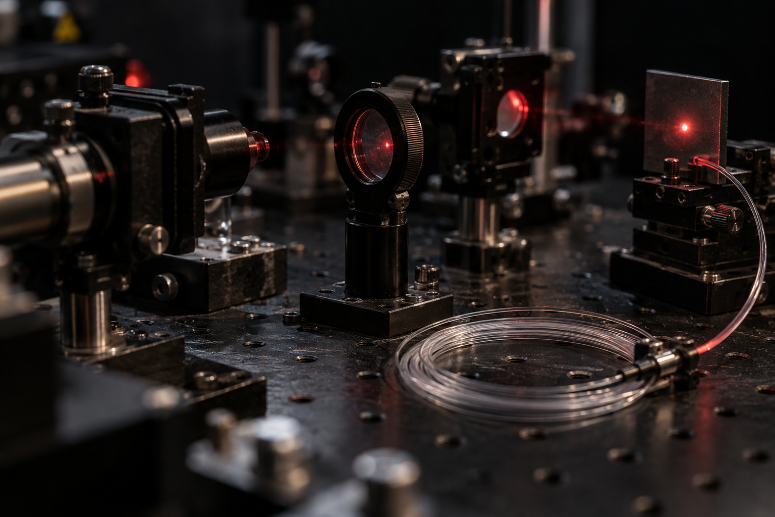
A laser’s risk is shaped by its wavelength, concentration, power, and exposure route. The eye can focus some beams onto delicate tissue, while ultraviolet wavelengths interact differently with skin and eyes; class labels summarize hazards but do not erase misuse risks.
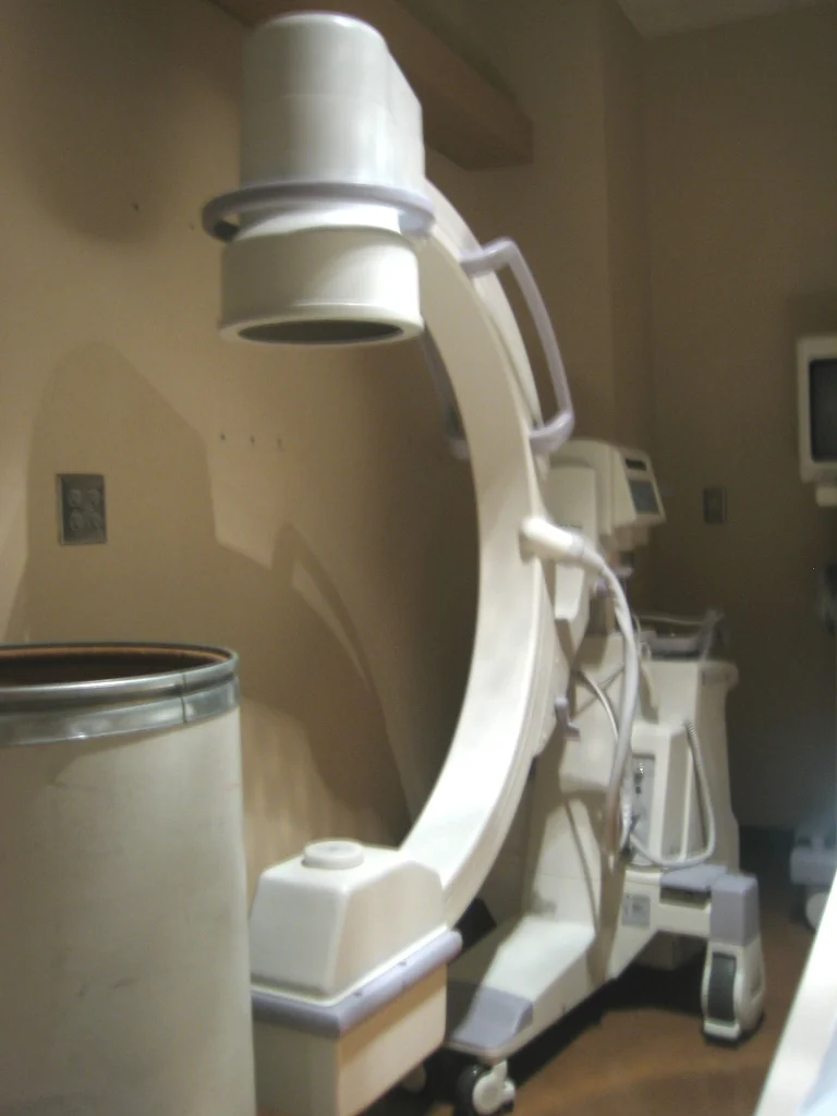
Fluoroscopy creates a sequence of X-ray images that lets clinicians watch anatomy, contrast material, or instruments move in real time. Its usefulness comes with a need to balance visibility against radiation exposure.
Cryosurgery uses a deliberately localized freeze to injure abnormal tissue. Its physical logic differs from heat-based hyperthermia, and the location, temperature pattern, and treatment purpose shape both effects and limits.
Fusion and medical plasmas share a state of matter but operate at radically different temperatures and for different purposes. Cold atmospheric plasma research explores surface interactions, with clinical evidence and appropriate uses still under evaluation.
A sensor’s electrical output becomes meaningful only when compared with known references. Calibration builds that bridge, while uncertainty and fitness for purpose determine how much confidence a measurement deserves.
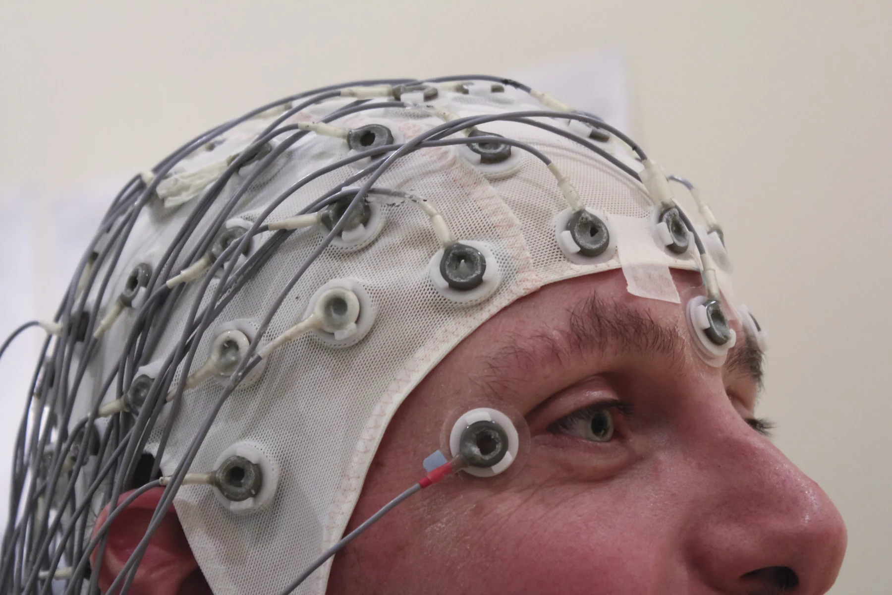
An EEG records voltage patterns at the scalp after signals from many brain cells pass through tissue and meet the outside world. That long path helps explain both the test’s strengths and its sensitivity to movement.
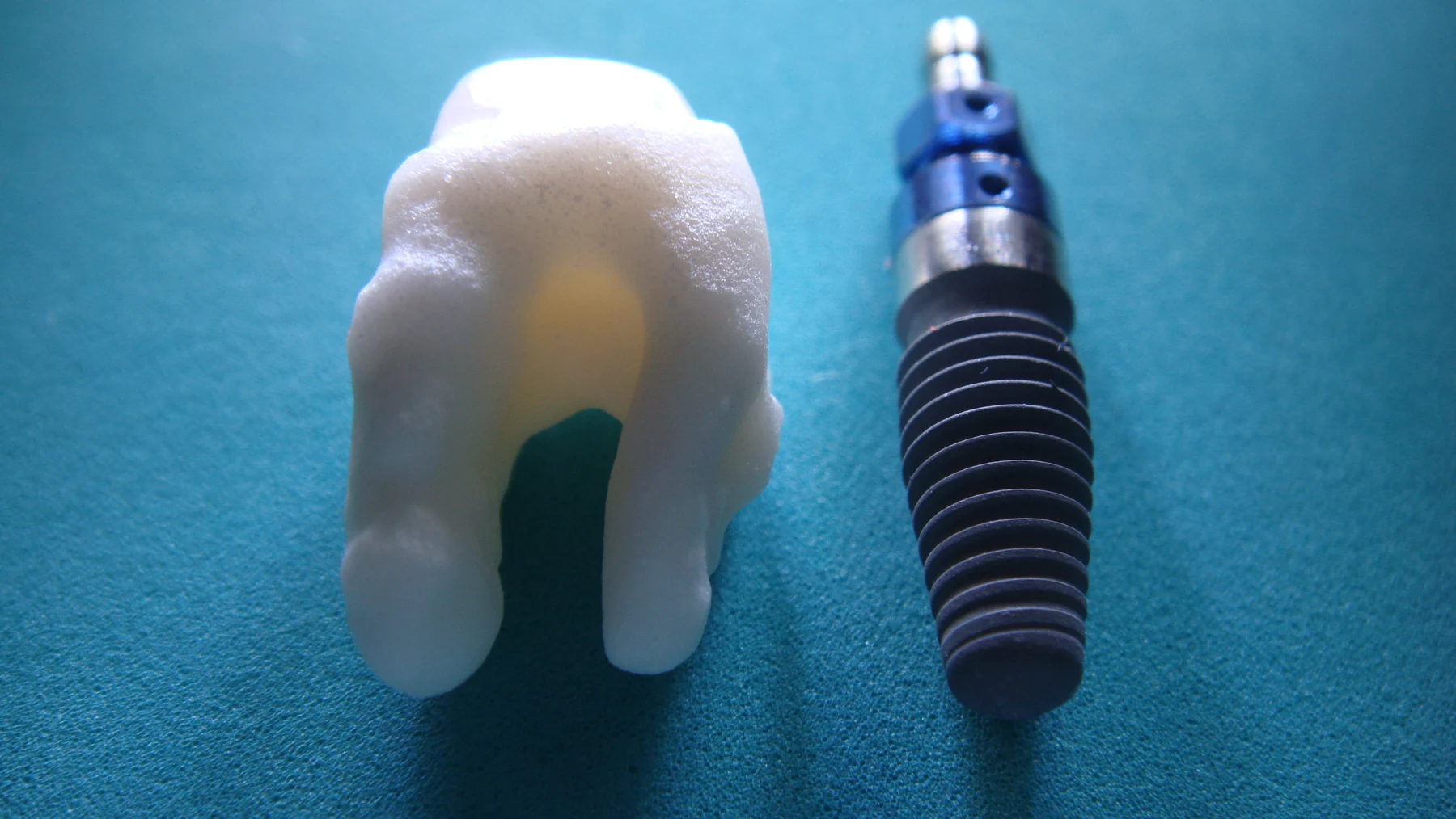
An implant can respond to MRI’s static and changing magnetic fields and radiofrequency energy, while also affecting the image. Identifying its exact status and conditions is part of safe, useful scanning.
Sterilization must inactivate microorganisms, yet the physical and chemical conditions used to do so can also affect a device. Material compatibility depends on the process, geometry, and intended use.
Radiation protection rests on three physical levers: reduce exposure duration, increase separation, and place suitable shielding in the path. Their use follows a more basic question: is the X-ray exam needed?
A material’s softness does not tell the whole story of how it bears force. Geometry, internal structure, and the way loads travel through it can make flexible biomaterials and robotic components useful supports.
A numerical difference can look encouraging yet remain uncertain, while a statistically detectable effect may matter little to patients. Reading clinical evidence requires attention to study design, uncertainty, outcomes, and the care standard being compared.
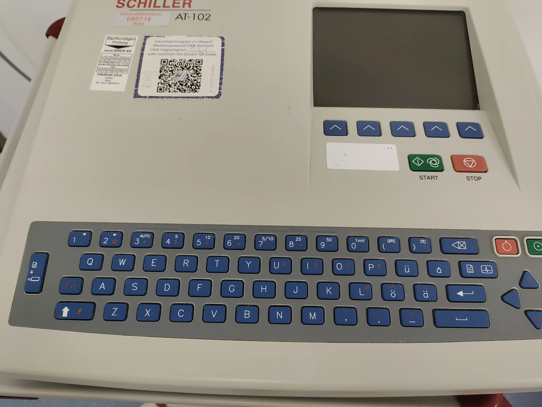
An ECG senses voltage differences at the skin as the heart’s electrical activation spreads and recovers. Its trace reveals timing and rhythm, but a short recording is only a sample of activity, not a diagnosis by itself.
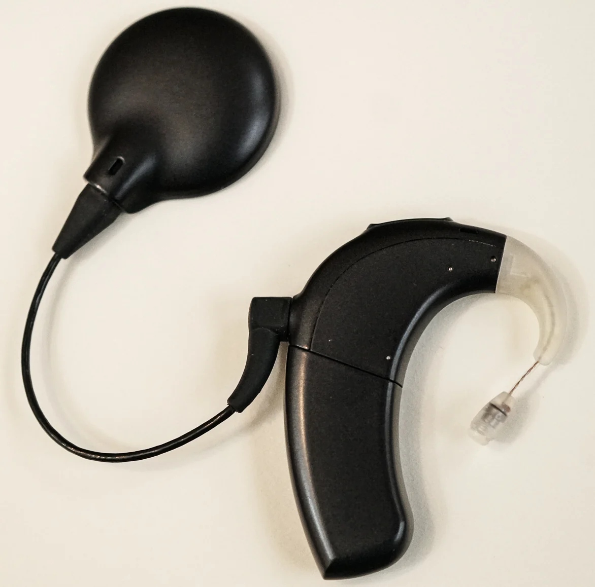
A cochlear implant converts sound into electrical signals and sends them to the auditory nerve, routing around damaged inner-ear hair cells. The result is a learned representation of sound, not normal hearing.
Brachytherapy treats a defined region by placing a radiation source in or near a tumor. Its geometry shapes dose distribution, while placement, timing, and nearby healthy tissue remain central to treatment planning.
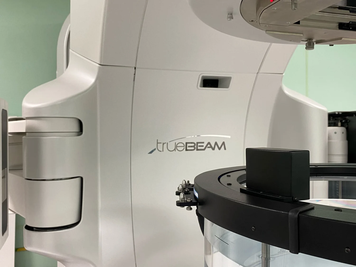
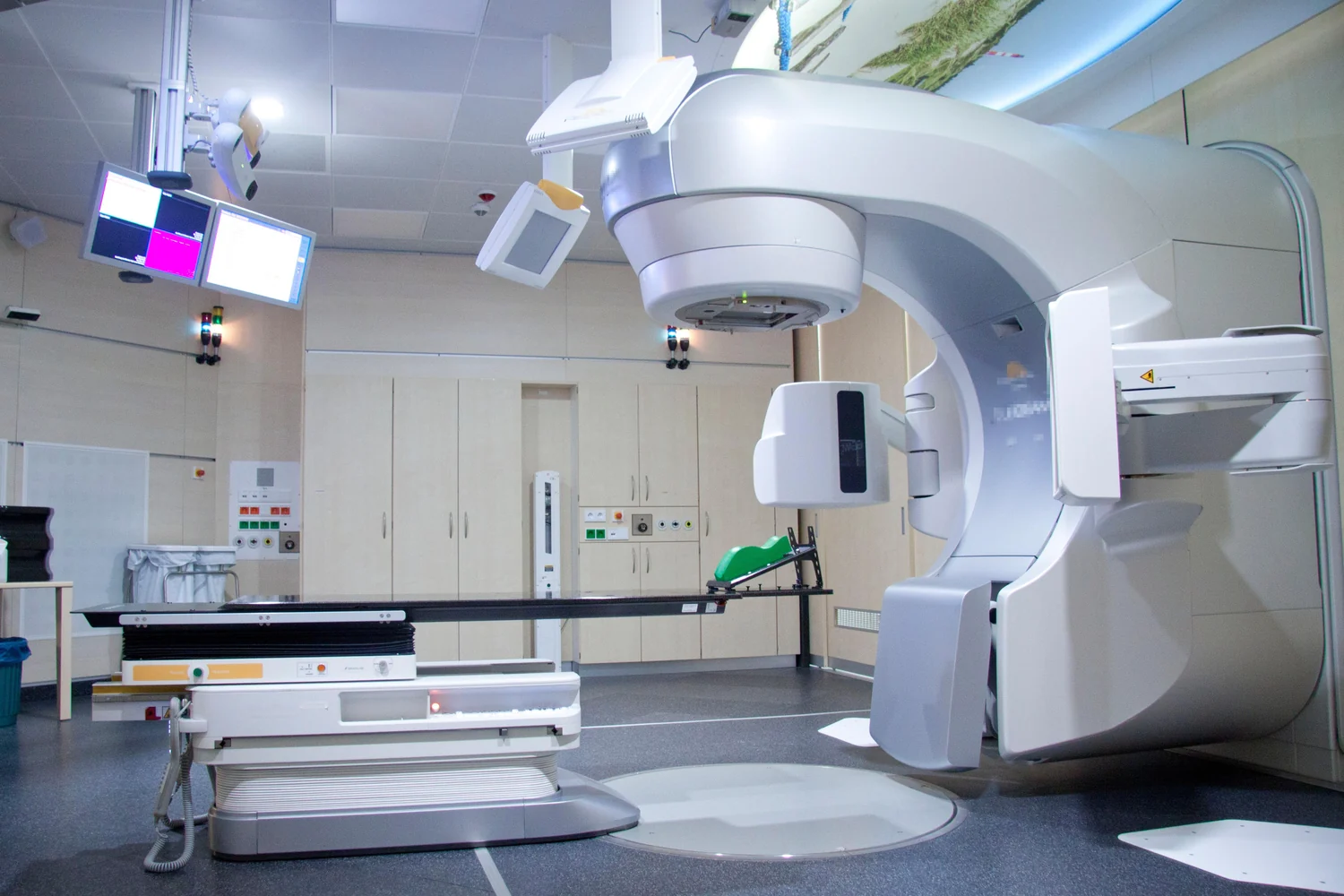
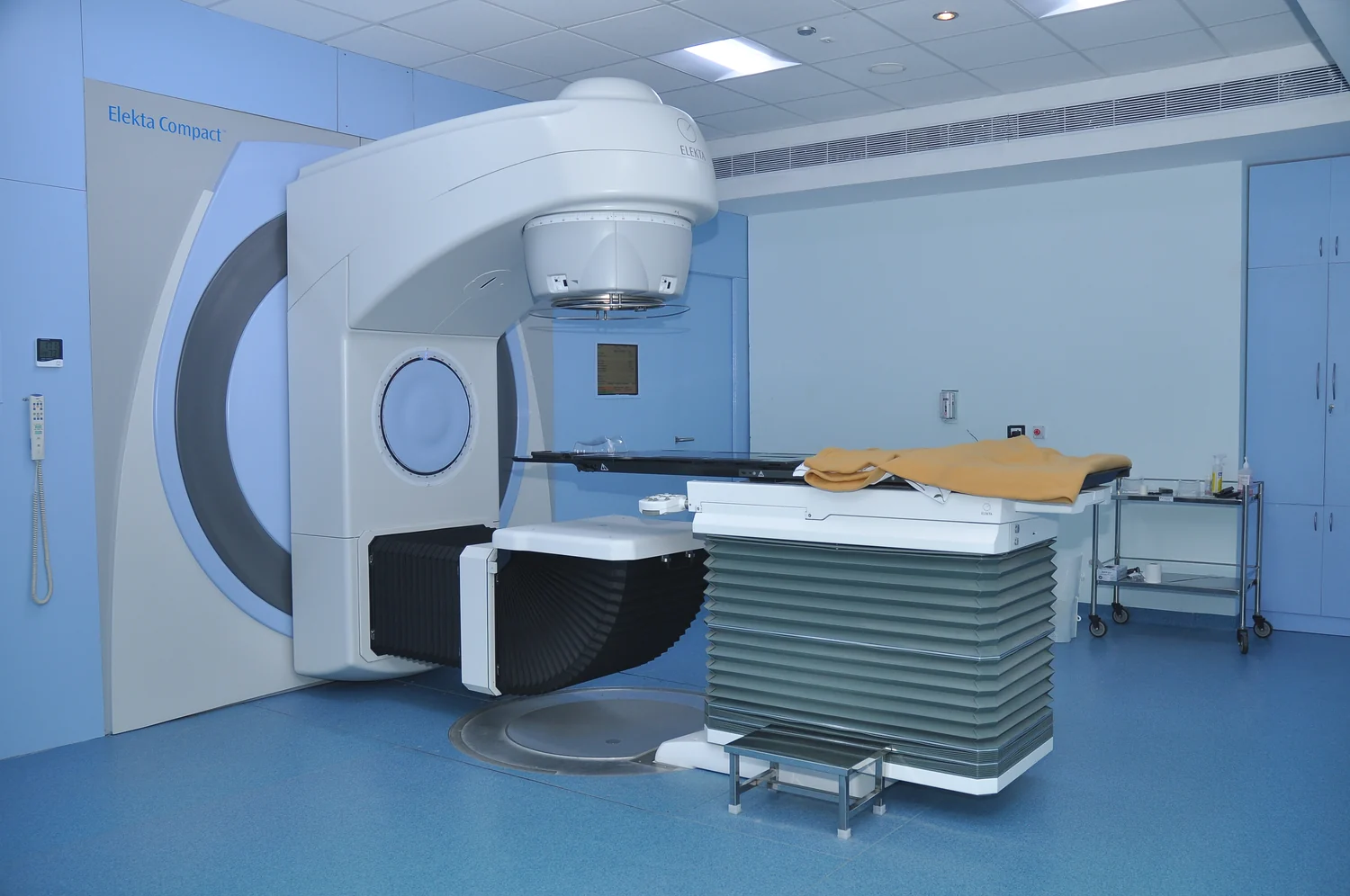
A linear accelerator helps shape and aim external radiation toward a planned target. Imaging, beam direction, and patient positioning work together to concentrate dose while limiting exposure to surrounding tissue.