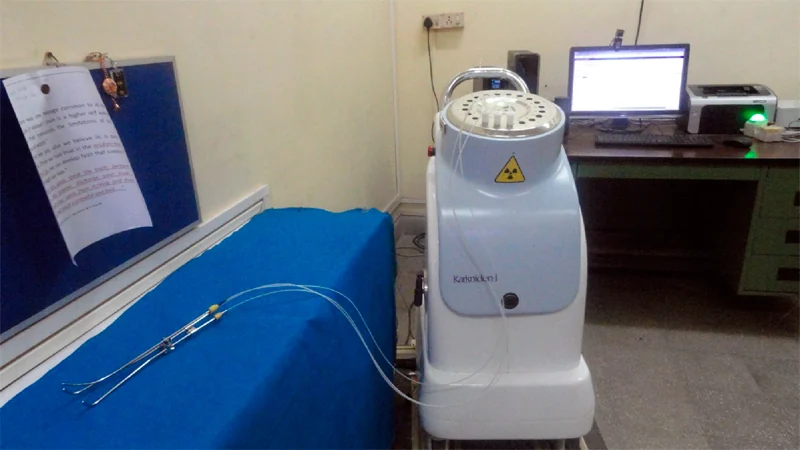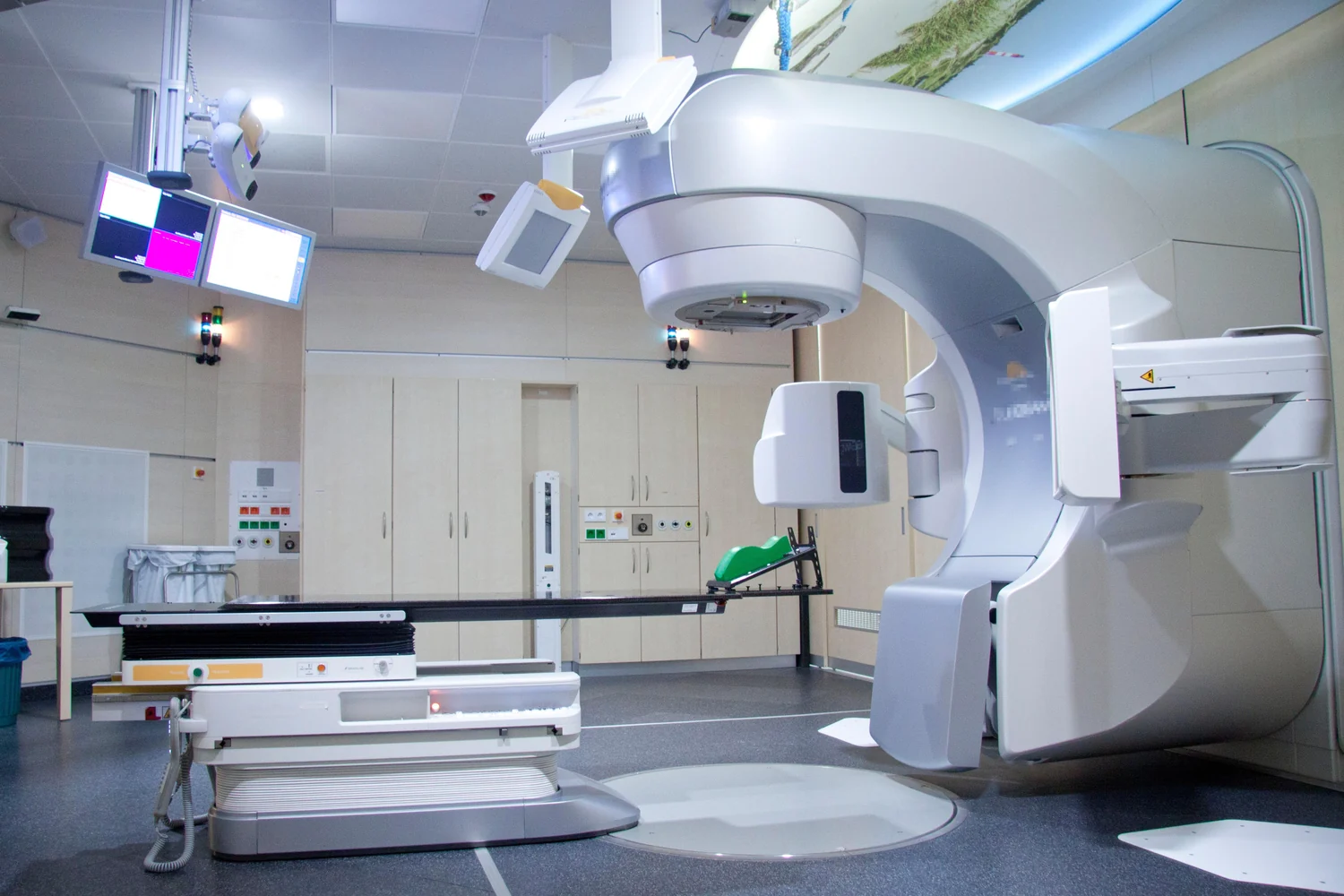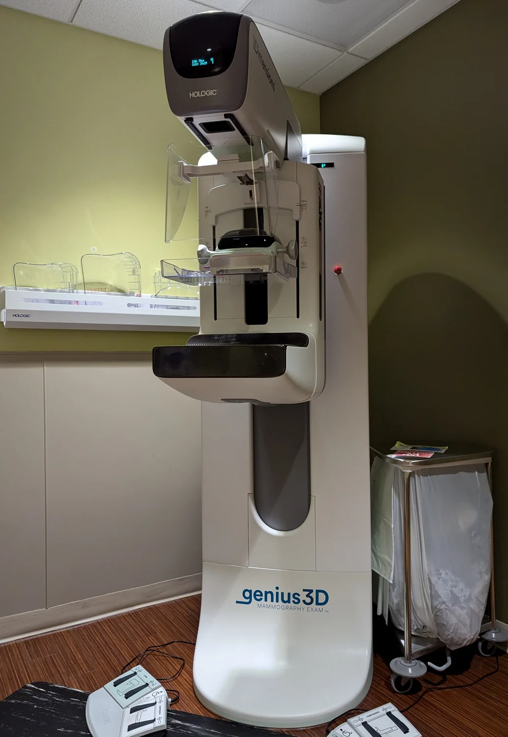
How can a room, a barrier, or a few steps change exposure to X-rays? The answer begins with radiation traveling from a source and interacting with matter. In medical imaging, protection is not a ritual of avoiding every photon; it is a way to keep exposure appropriate to the clinical task. Time, distance, and shielding help limit exposure, while the decision to image and the settings chosen determine whether the exam’s benefits justify its risks.
First ask why the beam is needed
An X-ray image begins when a beam passes through the body. Some photons are absorbed or scattered by internal structures; the pattern that remains reaches a detector. Radiography records an image, fluoroscopy displays a continuing image for real-time guidance, and CT combines many projections into cross-sectional slices. Each serves different questions, and CT generally involves more radiation than conventional radiography because it uses many projections [1].
The first protection principle is therefore justification: an exam using ionizing radiation should answer a medical question, help treat disease, or guide a procedure. Ionizing radiation has enough energy to damage DNA, and exposure can slightly increase the possibility of cancer later in life. For medically appropriate imaging, the expected clinical benefit generally outweighs the small risk; protection means considering that balance rather than treating all exposure as equally useful or harmful [2] [1].
Time: reduce unnecessary exposure
A simple mental experiment helps: imagine a person standing near a source while a beam is on. If the intensity and geometry stay the same, twice as much time in the radiation field means roughly twice the opportunity for exposure. Time matters especially in fluoroscopy, where images are displayed continuously while a team guides a procedure. Long, complex interventions can produce relatively high doses, including skin doses that may cause tissue effects in some circumstances [1].
This does not mean that a procedure should be rushed at the cost of a useful image or safe care. It means that the beam should be used for the needed task, and unnecessary exposure should be avoided. The optimization principle asks the imaging team to choose settings that provide image quality adequate for diagnosis or intervention, with dose as low as reasonably achievable. The patient’s size, the body area, and the clinical indication all affect those choices [1].
Distance: let space do some work
Radiation spreads as it travels away from a source. For a point-like source in open space, the same energy is distributed over a larger spherical area as distance increases; the intensity therefore falls approximately with the square of distance. Doubling the distance can reduce intensity to about one quarter under those simplified conditions. Real imaging rooms have scatter, barriers, and equipment, so this is a physical guide rather than a room-by-room dose calculation.
Distance is most useful for staff and others who do not need to remain close to the patient during an exposure. Moving away, where the procedure allows it, can reduce exposure from radiation scattered by the patient. The patient, however, is positioned to obtain the image or treatment guidance, so simply moving farther from the X-ray source is not a substitute for appropriate imaging technique. A clear clinical purpose and well-chosen equipment settings remain central [1].
Shielding: choose the right barrier
A shielding material reduces transmission by absorbing or scattering some of the radiation that reaches it. Dense materials such as lead are useful in X-ray protection because they interact strongly with X-rays. The barrier must be between the source of exposure and the person it is meant to protect, and its effectiveness depends on the radiation, material, thickness, and arrangement. A barrier is not a reason to expose an area without a clinical need.
The three levers work together, but they do not replace judgment. FDA guidance emphasizes justification and optimization, including equipment settings that produce an adequate image at the lowest reasonable dose. Risk also depends on factors such as age, body region, and dose; younger patients are more sensitive, which is why pediatric imaging receives particular care. In practice, protection is a design problem: obtain information that can change care while limiting exposure that contributes nothing to the answer [1] [2].
Sources & further reading
- Medical X-ray Imaging FDA
- Radiation National Cancer Institute
Prepared for the independent PSI Center reading library. About edition dates and source checking.
This explainer describes scientific principles. It does not assess individual treatment choices.