Why can the same broad kind of radiation be used to make an image and to treat cancer? The answer lies in the job being asked of it. A diagnostic scan needs enough x-ray information for a computer to distinguish structures. Radiotherapy deliberately delivers a much higher dose to damage cancer-cell DNA, with planning aimed at controlling where that energy goes.
An image needs contrast
In an x-ray examination, radiation passes through the body, and tissues absorb or scatter different amounts. A detector records the pattern that emerges. Conventional radiography captures a projection; CT collects many projections from different angles and reconstructs cross-sectional slices. The useful signal is the difference among tissues, not injury to them. [1]
Every x-ray exposure carries some ionizing radiation, which has enough energy to potentially damage DNA. For most medically appropriate imaging, the risk of a later cancer is small, and the examination is chosen because the information can help answer a medical question or guide care. CT typically uses a higher dose than a single radiograph because its image is reconstructed from many x-ray views. [1]
Treatment seeks biological damage
Radiotherapy uses high doses of radiation to damage the DNA of cancer cells. If the damage is beyond repair, cells stop dividing or die; the body then breaks down and removes them. This process is not instantaneous. The effects can build over days or weeks of treatment and continue for weeks or months after treatment ends. [2]
A useful distinction is between sensing and changing. In imaging, the beam is a probe: the desired result is a readable pattern at the detector. In radiotherapy, energy is the intervention: the desired result is enough damage in a target to control disease. The term radiation covers both uses, but their purpose and dose scale differ substantially.
Aim the energy; protect what surrounds it
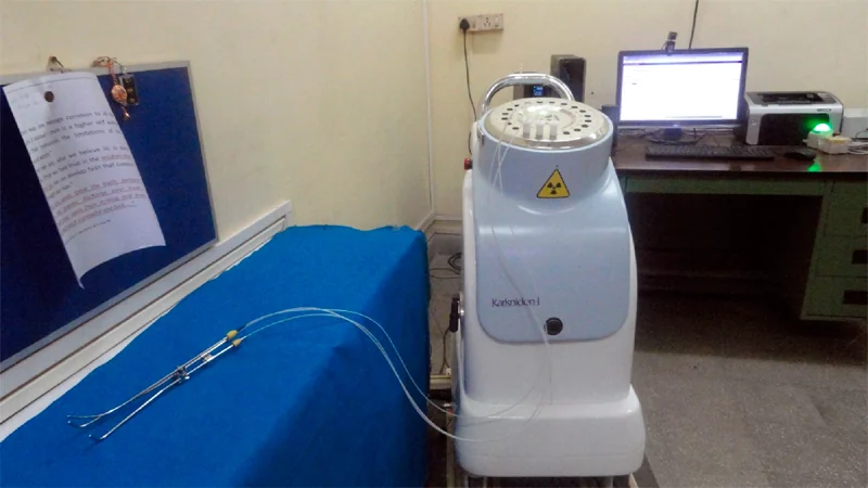
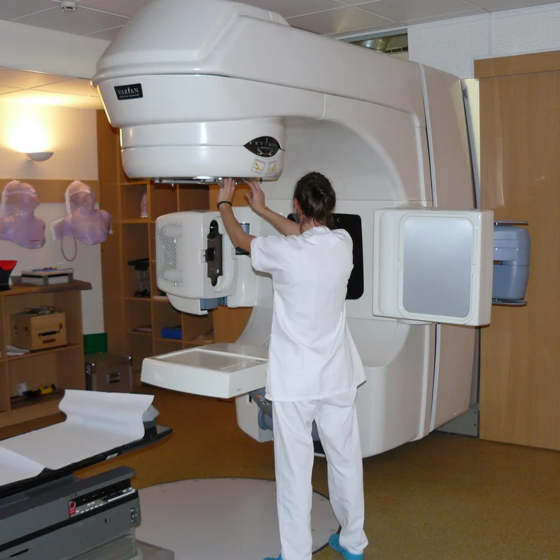
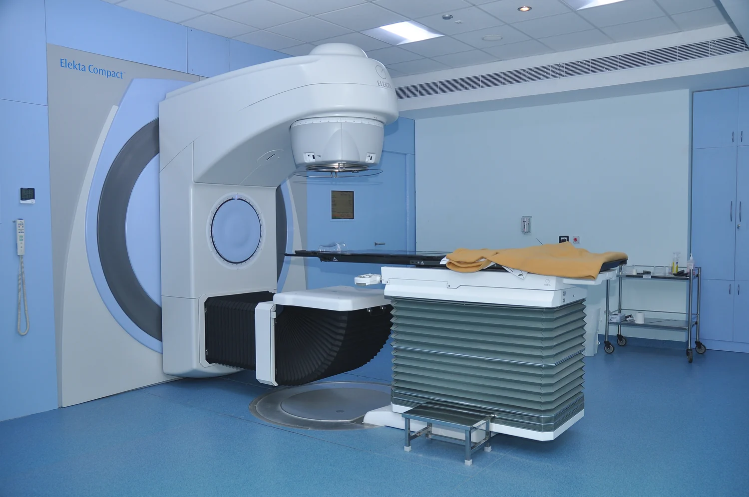
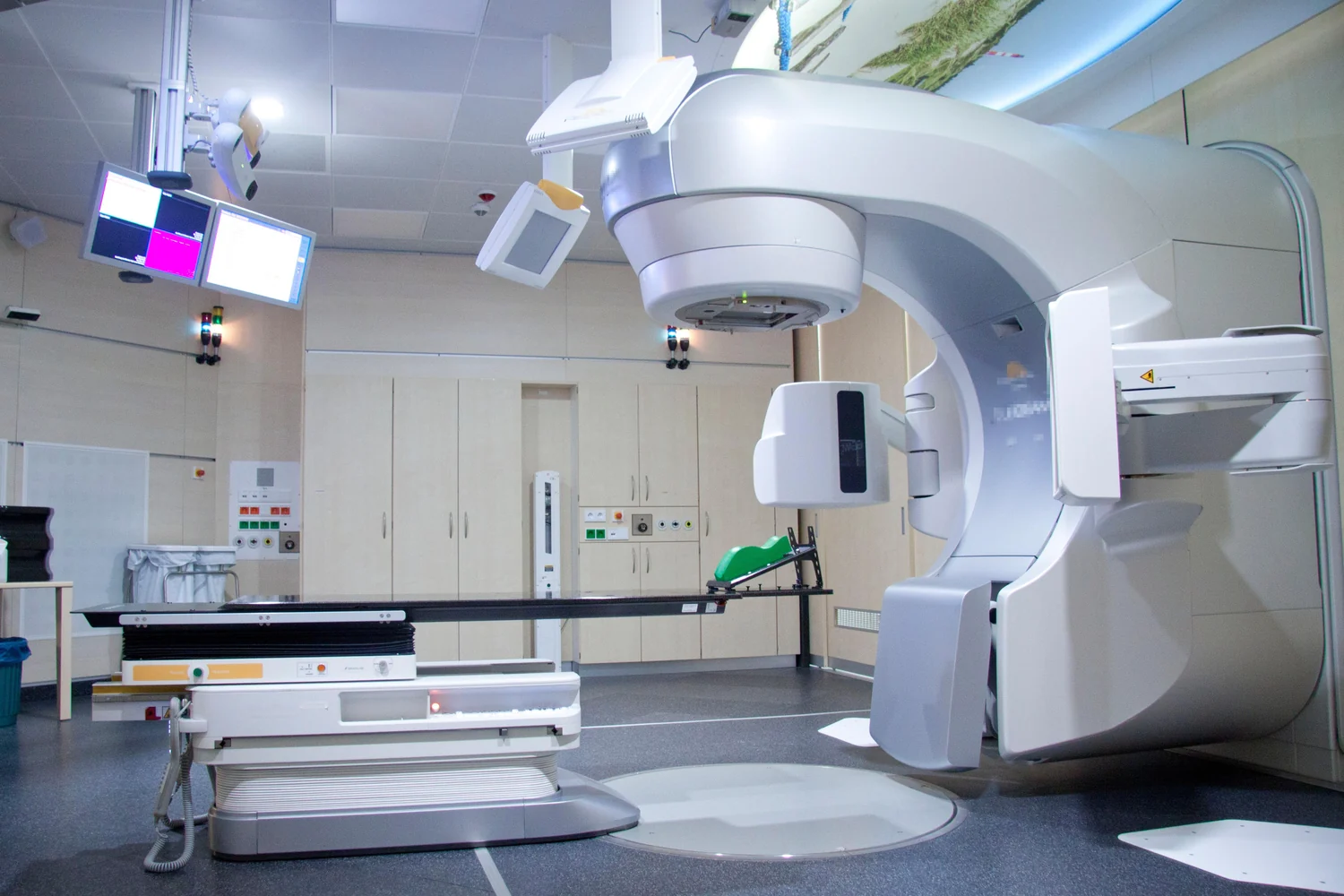
External-beam radiotherapy comes from a machine that directs radiation toward a selected region. It can approach a target from multiple directions, helping shape where dose accumulates. Internal methods place a radiation source in or near a tumor, or deliver radioactive material through the body in selected therapies. Which approach fits depends on the cancer, its location, and nearby sensitive tissues. [2]
Neither method can perfectly separate a tumor from healthy biology. Radiation may affect nearby healthy cells, causing side effects. Treatment planning therefore balances the dose needed for cancer control with the exposure of surrounding tissue. Radiotherapy is generally local when delivered as external beam or brachytherapy, while some systemic radioactive treatments travel through the bloodstream.
Different safeguards for different goals
For diagnostic imaging, justification means using an x-ray exam when it can do more good than harm for the clinical question. Optimization means adjusting the technique to use the lowest dose that still provides adequate image quality. These principles do not mean making every image as faint as possible: an image that cannot answer the question may lead to repeat imaging or missed information. [1]
Radiotherapy has its own planning problem: deliver a therapeutic dose to the intended target while limiting exposure to normal tissue. A treated body region also has a lifetime tolerance, so repeating radiation to the same area can be constrained; another sufficiently separate area may still be considered. In both settings physics serves medicine, but the success criteria differ. One aims for a useful signal with minimal exposure; the other aims for controlled biological injury where treatment is needed. [2]
Sources & further reading
- Medical X-ray Imaging FDA
- Radiation Therapy to Treat Cancer National Cancer Institute
Prepared for the independent PSI Center reading library. About edition dates and source checking.
This explainer describes scientific principles. It does not assess individual treatment choices.