Why does an EEG need a set of electrodes spread across the scalp to record what brain cells are doing? The electrical activity is real, but the signals measured at the skin are small and mixed together. They have traveled through brain tissue, fluid, skull, and scalp, while movement and muscle activity can add their own voltage changes. Understanding that path helps explain why an EEG is useful as a pattern-reading tool, yet easily disturbed by ordinary motion.
From many cells to one scalp electrode
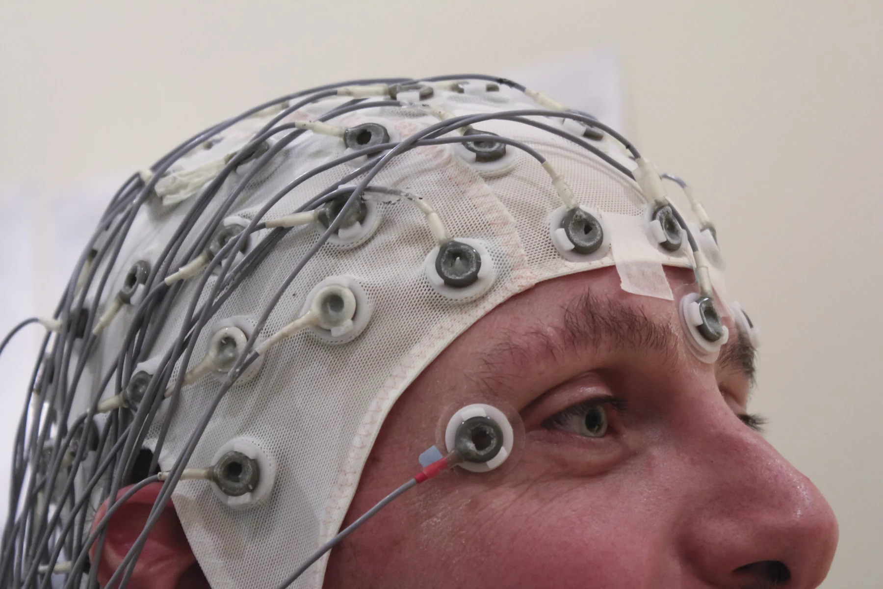
Brain cells communicate through tiny electrical impulses. An electroencephalogram, or EEG, measures patterns of that activity using flat metal electrodes placed at many points on the scalp. The recording machine converts the signals into wave-like traces on a monitor or paper. It is not listening to a single neuron; each electrode samples a blended electrical field produced by activity across groups of cells. [1]
A helpful mental picture is watching a stadium from outside through several walls. A coordinated crowd can create a broad rhythm, but an observer cannot pick out each person’s voice. In the same way, electrical signals from the brain must pass through intervening tissues before reaching the scalp. The EEG’s strength comes from recording changing patterns across locations and time, not from isolating every cell or pinpointing every event to a single source.
The path weakens and mixes the signal
Electrical currents spread through conductive tissue rather than traveling like a narrow wire from one cell to one sensor. As activity passes through brain and surrounding tissues, the signal reaching a scalp electrode is spatially blurred and relatively small. Each electrode also receives contributions from multiple sources. This mixing is one reason EEG interpretation focuses on organized wave patterns and their timing, rather than treating a scalp trace as a direct readout from one brain structure. [1]
The setup is designed to preserve useful contact. Electrodes are held in place with sticky paste, and preparation instructions may include washing hair and avoiding products that interfere with that contact. The person is asked to lie still because motion changes the recording. A blink, a jaw clench, or a shifted electrode can alter the voltage at the sensor. Such changes may be larger than the brain signal of interest, much as a nearby drumbeat can drown out a quiet conversation. [1]
Muscles are electrical neighbors
Muscle activity is not merely mechanical movement; contracting muscle produces electrical activity too. An electromyography test, or EMG, measures that activity with a thin needle electrode placed in a muscle. The electrical pattern changes when the person contracts the muscle, and it can help distinguish weakness related to nerve injury from weakness caused by muscle or nervous-system disease. [2]
That contrast clarifies why facial and neck tension can complicate an EEG. The brain and muscles share the same recording environment, and electrical signals from active muscles can reach scalp electrodes. Even a person trying to remain still may tense muscles without noticing. EEG staff may therefore repeat or compare portions of a trace and use the circumstances of a signal change to assess whether it reflects brain activity or interference. A waveform is meaningful only in context, including what the person was doing at the time. [1] [2]
Patterns, not a simple brain meter
An EEG is used to diagnose or monitor conditions such as seizures and epilepsy, and it can also be used in sleep evaluation or during brain surgery. Normal patterns vary with alertness: waves are faster during wakefulness and slower in some stages of sleep. The recording therefore needs to be interpreted against state and timing. A normal result does not prove that a seizure never occurred; it means that the recorded sample did not show a diagnostic pattern. [1]
Some procedures deliberately ask a person to breathe rapidly and deeply or look at a flashing light because these conditions can change brain activity; in people with seizure disorders they may trigger a seizure, so trained personnel supervise the test. Longer ambulatory recordings can capture activity over time during a person’s routine, sometimes with video to relate the signal to behavior. The measurement is a window into electrical patterns, not a direct measure of intelligence, and not a complete account of everything the brain is doing. [1]
Sources & further reading
- Electroencephalogram MedlinePlus
- Electromyography MedlinePlus
Prepared for the independent PSI Center reading library. About edition dates and source checking.
This explainer describes scientific principles. It does not assess individual treatment choices.