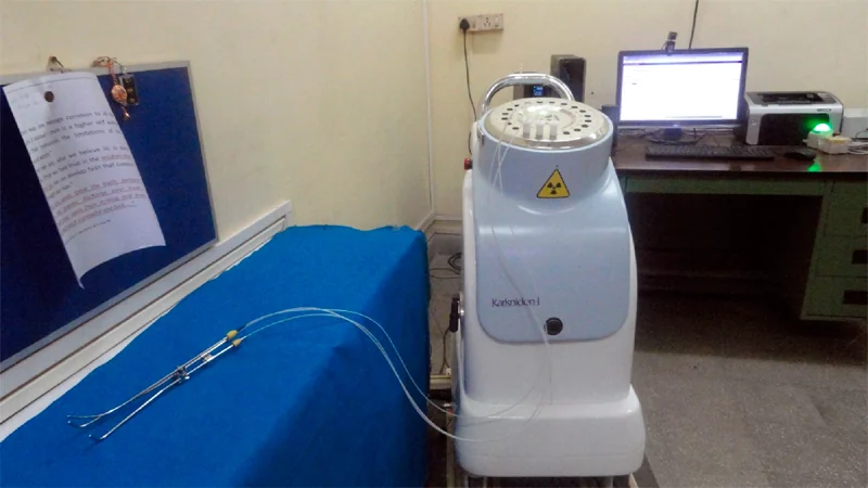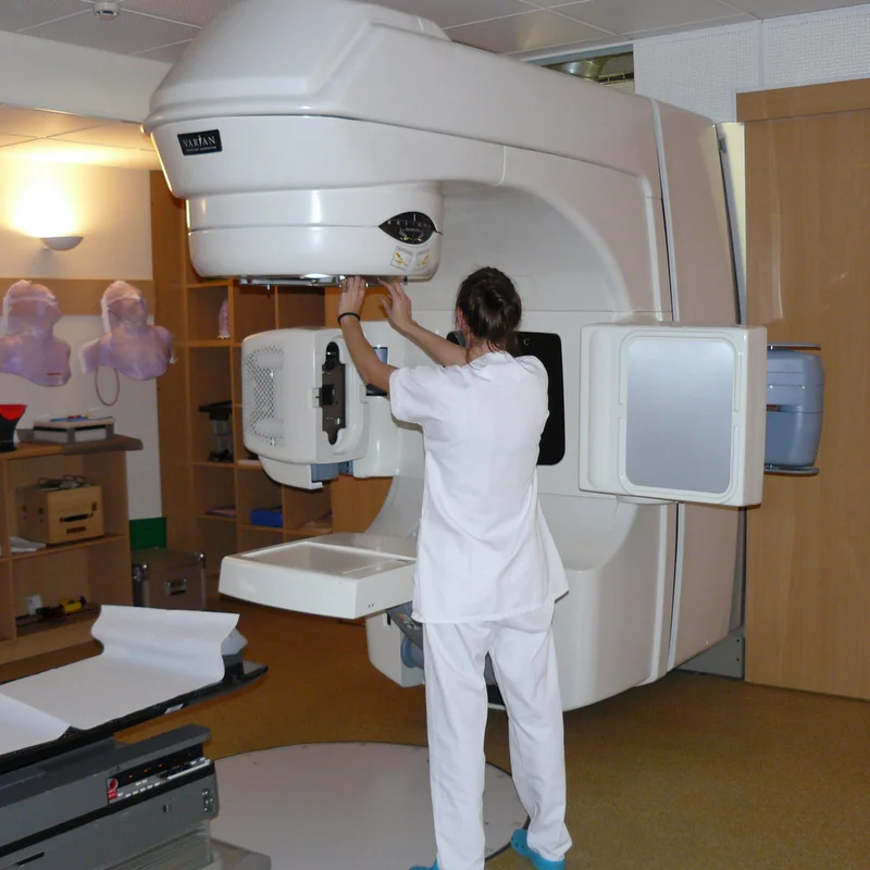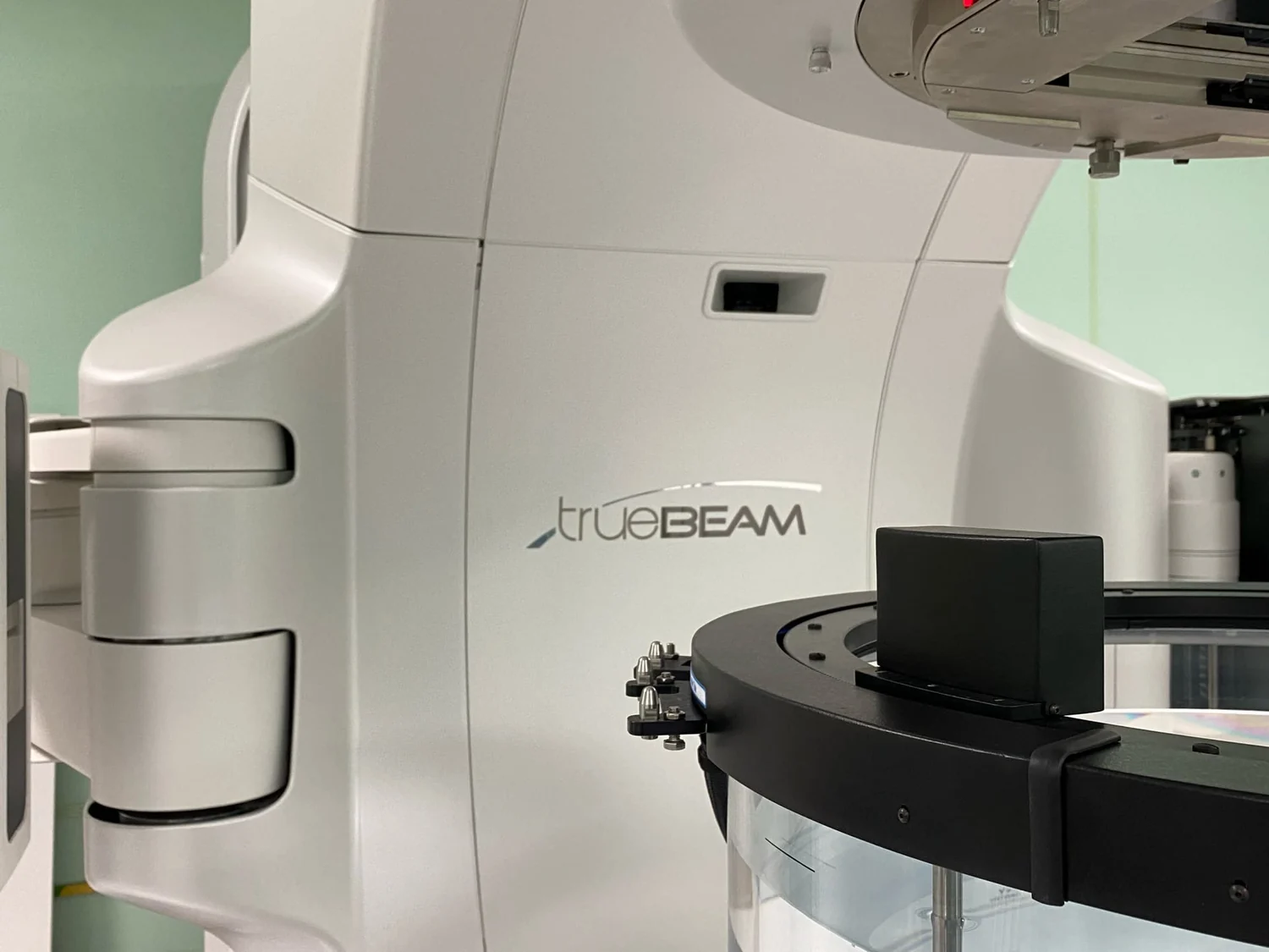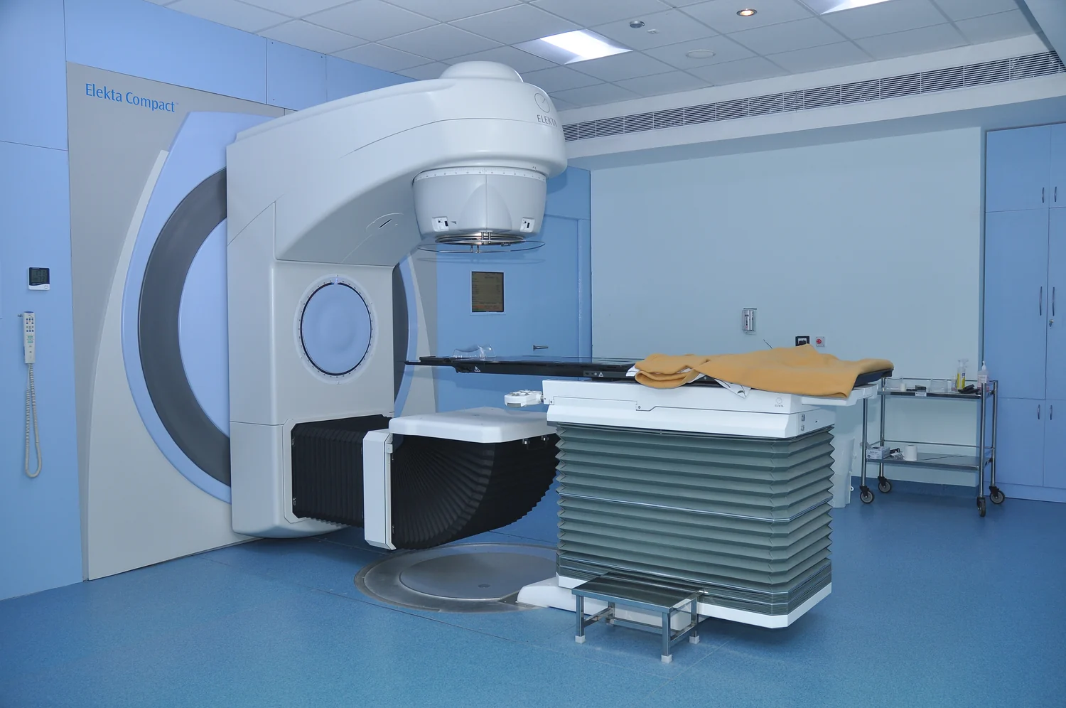
How can a machine outside the body deliver radiation to a tumor inside it? A linear accelerator produces a beam that can be directed toward a planned treatment area. The machine’s contribution is not simply generating radiation: imaging, dose calculation, beam shaping, and reproducible positioning help determine where energy is deposited and how much nearby tissue is exposed.
What the machine sends
External beam radiation therapy uses a machine to aim radiation at a specific part of the body. Many machines use photon beams, which can reach deep tumors but also scatter some radiation along their path and continue beyond the target. Other external beams use protons or electrons, each with different physical properties and suitable depths. A linear accelerator is one kind of machine used to produce treatment beams. [1]
Radiation damages cancer-cell DNA. If the damage is beyond repair, cells stop dividing or die, but that process takes time: effects can unfold over days and weeks, including after treatment ends. The beam is therefore not a visible projectile that instantly removes a tumor. It deposits energy that initiates biological changes in cells, while treatment planning aims to place the prescribed dose in the intended region. [2]
Planning the path through the body
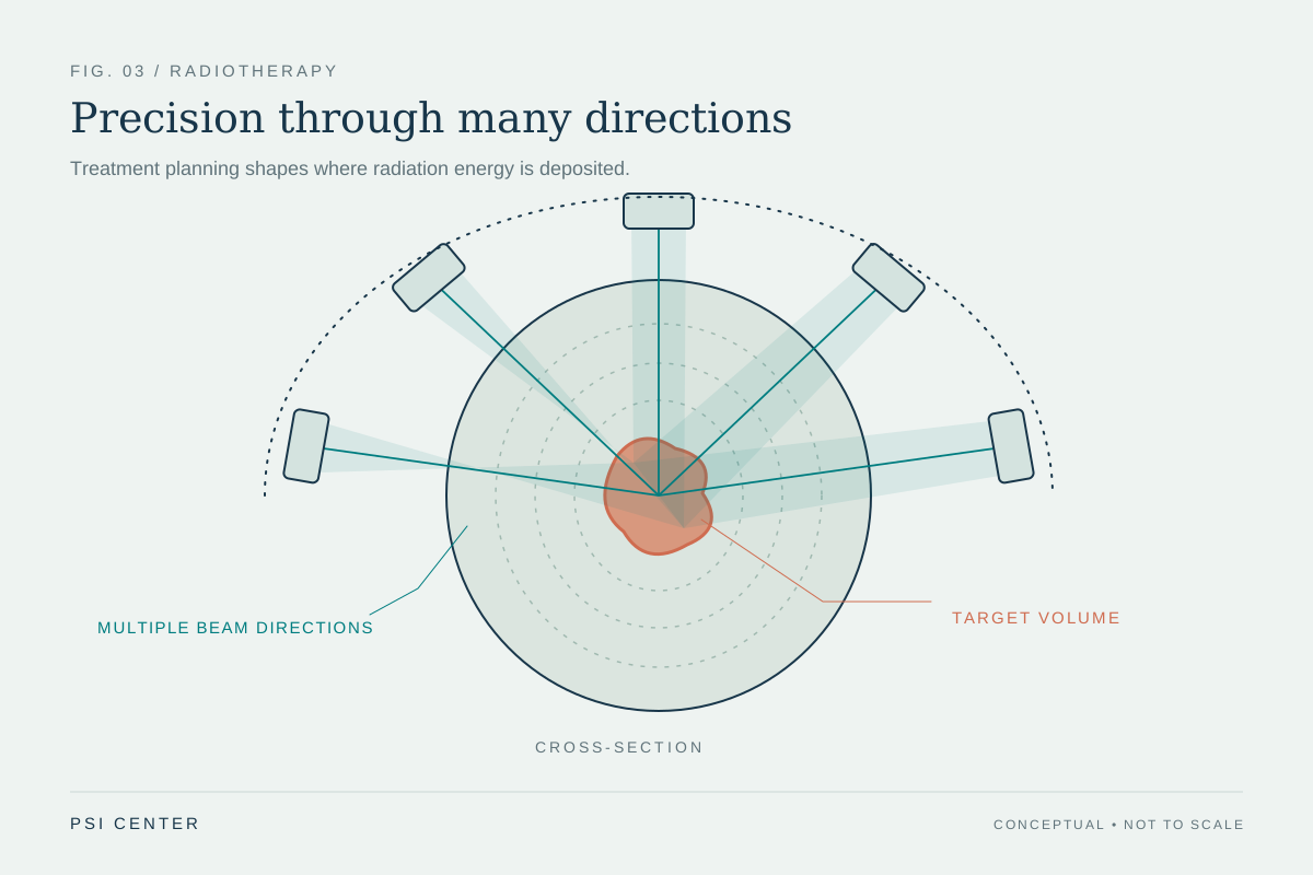
Before treatment, imaging helps define the target and its relationship to nearby structures. In three-dimensional conformal therapy, CT, MRI, or PET images inform a computer plan, and beams approach from multiple directions. Intensity-modulated radiation therapy uses many smaller beams whose strength can vary across the field, allowing different parts of the tumor to receive different intensities. [1]
The geometry matters because each beam travels through tissue. With multiple directions, the planned contributions overlap at the target, while each path may deliver a smaller portion of the total dose to other locations. This is a useful mental model for focused lighting: several dim beams can illuminate one spot more strongly where they converge. Real radiation planning is more complex, and the dose distribution depends on anatomy, beam type, and the treatment plan.
Repeatable position, repeatable aim
A plan works only if the patient and machine are aligned as expected. During simulation, the treatment area is mapped and marks, molds, or masks may help reproduce the same position at each session. Image-guided radiation therapy adds scans during treatment sessions; computer processing can detect changes in tumor size or location and support adjustments to position or dose. [1]
This is why a session may involve more time for setup than for beam delivery itself. The NCI describes treatment beams being delivered for minutes, while visits often last longer because positioning must be checked. The machine contributes precision through controlled motion and beam delivery, but staff, imaging, and immobilization methods are part of the system that makes the planned geometry possible. [1]
Precision has limits and tradeoffs
Radiation is local in the sense that external beam therapy treats a specific region, but nearby healthy cells can also be affected. That can cause side effects. The aim across different external beam methods is to deliver the prescribed dose to the tumor while sparing normal tissue as much as possible; no beam geometry makes the body’s surrounding tissues disappear from the problem. [2] [1]
Treatment schedules vary with the cancer, target, and goal. Many people receive sessions on weekdays over a course of treatment, allowing time between doses for healthy cells to recover and making radiation more effective. A linear accelerator thus contributes a controllable source and delivery geometry. It does not determine by itself who should receive treatment, what schedule is suitable, or whether radiation is used alone or with other care. [1]
Explore the physics: an explanatory diagram
Sources & further reading
- External Beam Radiation Therapy National Cancer Institute
- Radiation Therapy to Treat Cancer National Cancer Institute
Prepared for the independent PSI Center reading library. About edition dates and source checking.
This explainer describes scientific principles. It does not assess individual treatment choices.