Why Plans Need Independent Checks
A treatment plan depends on measurements, images, calculations, and patient positioning. Independent checks help catch errors and test whether the planned delivery matches the intended target and constraints.
A closer look at the science
behind better healthcare.
How radiation is measured, shaped, and used to treat cancer, with clear explanations of benefits and limits.
41 articles
Clear filtersA treatment plan depends on measurements, images, calculations, and patient positioning. Independent checks help catch errors and test whether the planned delivery matches the intended target and constraints.
Systemic radiation therapy uses a carrier molecule to bring radioactive atoms through the bloodstream toward selected cells. Its reach can include many sites, while targeting and nearby-tissue exposure remain central constraints.
Protons lose energy as they pass through tissue, with energy deposition rising near the end of their path. The Bragg peak helps explain proton therapy’s potential precision and why range uncertainty still matters.
Radiation planning turns scans, beam properties, and a prescribed target into a calculated treatment plan. Modeling helps estimate where energy will go while accounting for the surrounding anatomy.
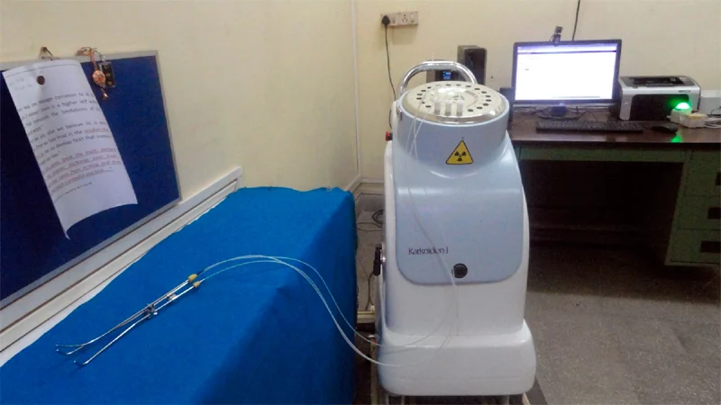
Brachytherapy places a radiation source in or near a tumor. Because radiation intensity and tissue exposure vary with geometry, source placement is central to delivering a local treatment while limiting exposure beyond the target.
Proton therapy differs from photon radiation in how energy is deposited along a beam’s path. That physical distinction can help shape treatment, while clinical benefit depends on tumor geometry, nearby organs, and evidence for each cancer.
Cancer research moves through questions of mechanism, safety, and clinical benefit. Keeping those stages distinct helps explain what a promising result can—and cannot—tell patients.
Radiation treatment depends on delivering a planned dose to a defined target while limiting exposure to nearby tissue. Measurement chains and repeated imaging help make those plans interpretable and checkable.
Gray measures energy deposited per unit mass; sievert expresses radiation dose with biological-effect weighting. The units answer different questions, so choosing between them depends on whether the concern is energy absorption or protection.
Brachytherapy places radiation close to a target, but the source’s residence time changes both treatment logistics and the radiation around it. Understanding that distinction clarifies why temporary and permanent implants call for different safeguards.
Photon and electron beams are both used in external radiation therapy, but they travel through tissue differently. Their paths help explain why treatment planning aims to place dose in a target while limiting exposure nearby.
Cryosurgery uses a deliberately localized freeze to injure abnormal tissue. Its physical logic differs from heat-based hyperthermia, and the location, temperature pattern, and treatment purpose shape both effects and limits.
Radiation protection rests on three physical levers: reduce exposure duration, increase separation, and place suitable shielding in the path. Their use follows a more basic question: is the X-ray exam needed?
Brachytherapy treats a defined region by placing a radiation source in or near a tumor. Its geometry shapes dose distribution, while placement, timing, and nearby healthy tissue remain central to treatment planning.
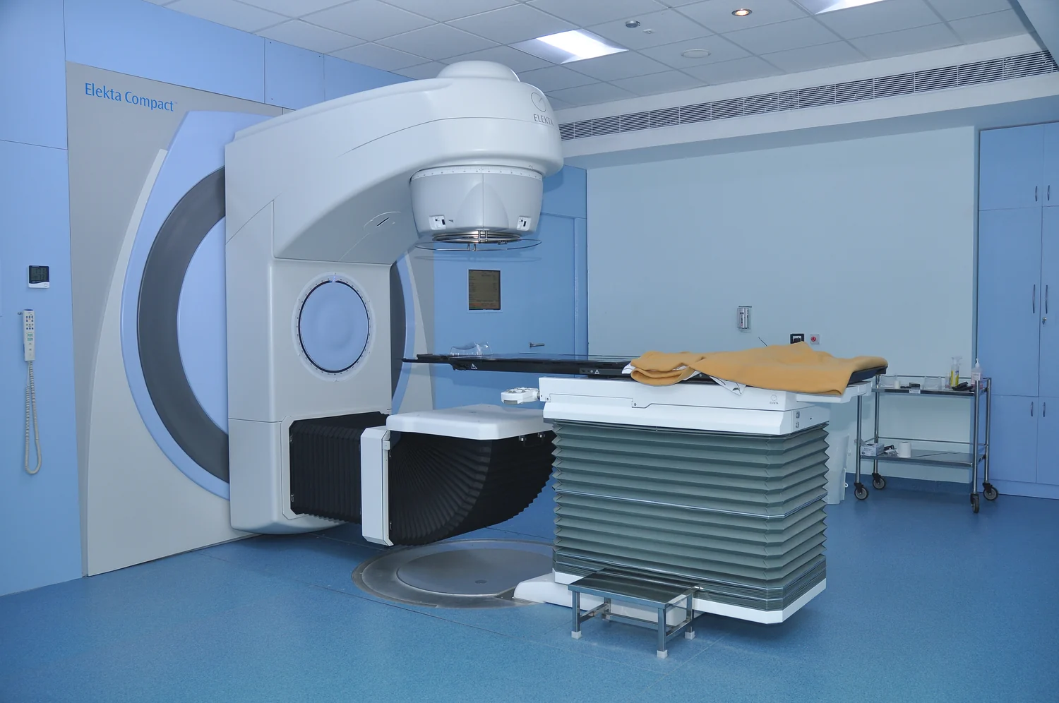
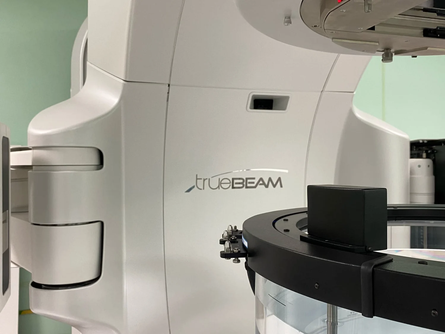
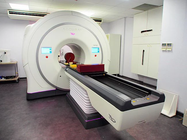
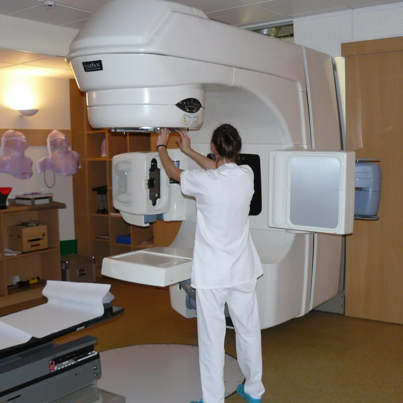
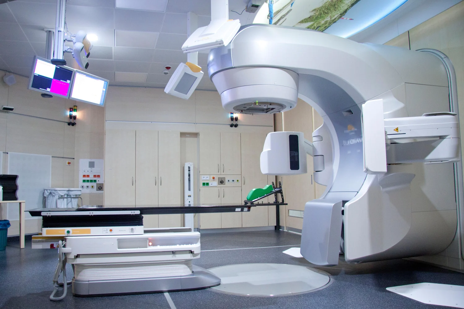
A linear accelerator helps shape and aim external radiation toward a planned target. Imaging, beam direction, and patient positioning work together to concentrate dose while limiting exposure to surrounding tissue.
Cancer hyperthermia aims to use heat alongside other treatments, but the temperature inside a tumor can vary from nearby tissue. Probes, imaging, and models help explain why careful control matters.
Imaging and radiation treatment both use ionizing radiation, but they pursue different physical goals. A scan seeks enough signal to answer a question; therapy aims to damage tumor-cell DNA while limiting harm to nearby tissue.
Radiation plans rely on knowing where a target sits, but tumors in the chest and abdomen can move as a person breathes. Imaging and computational models help teams account for that changing position.