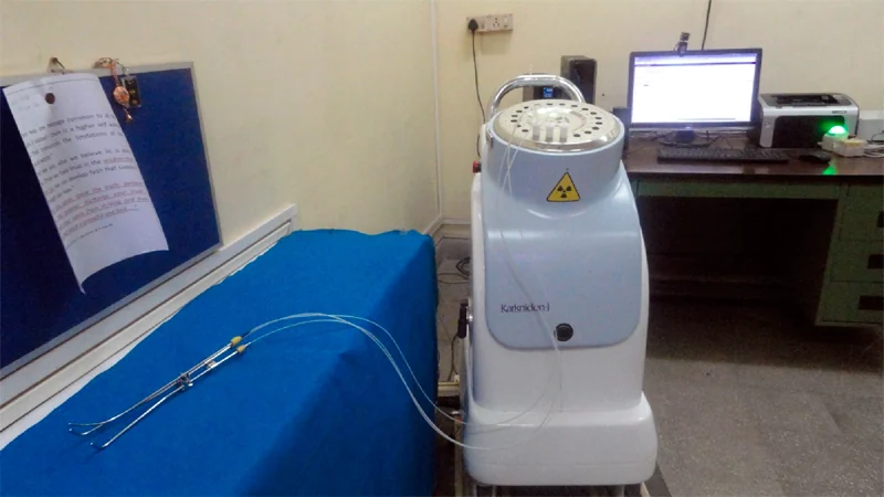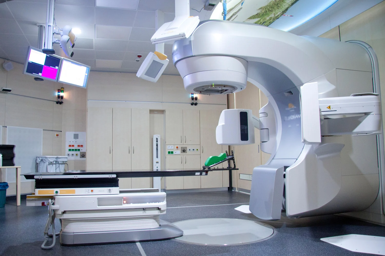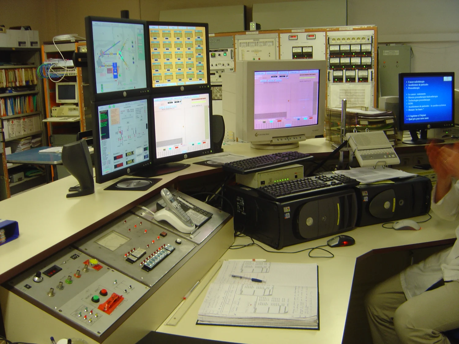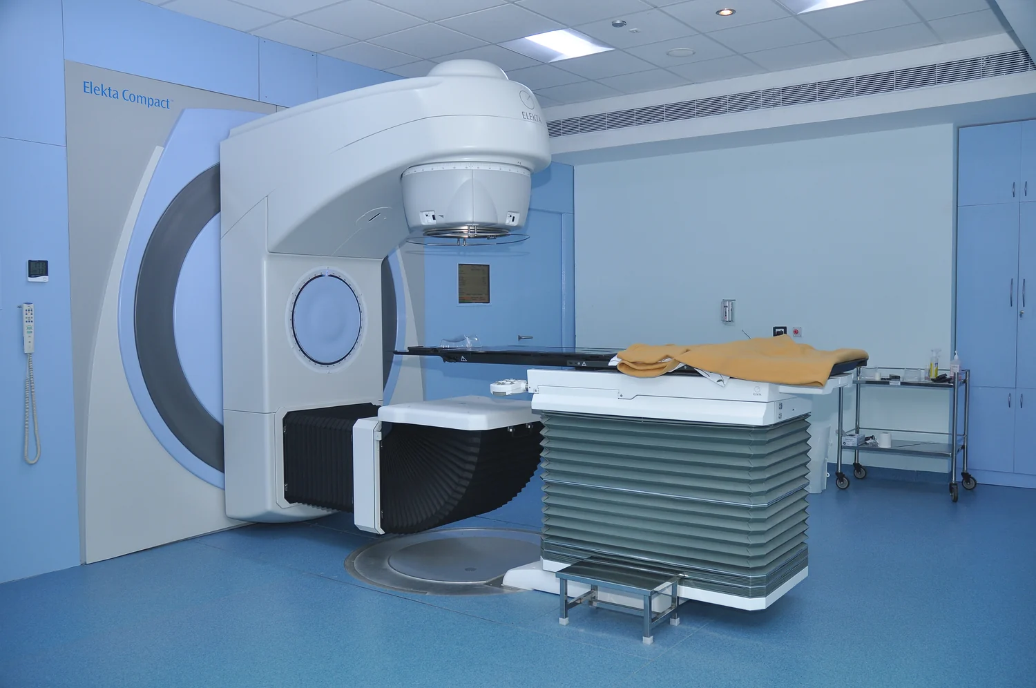
Why might a radiation machine use photons for a tumor deep in the body but electrons for one near the skin? The particles in the beam follow different paths through tissue. Photons can penetrate deeply and continue beyond a target, while electrons do not travel as far. Those differences influence how clinicians plan external-beam treatment, whose purpose is to deliver a prescribed dose to a selected region while limiting dose to surrounding normal tissue.
What the beam carries
Photons are packets of electromagnetic radiation. In external-beam radiation therapy, they are used by most treatment machines and can reach tumors deep within the body. X-rays are also photons, though diagnostic X-ray imaging uses lower doses than radiation therapy. Photons transfer energy as they interact with tissue; along their route, some radiation is deposited in healthy tissue as well as in the intended target. [1]
Electrons are particles with negative electric charge. They interact with matter differently and cannot travel as far through body tissues as photon beams. That limited penetration makes electron beams useful for tumors at or near the surface, but unsuitable for reaching many deep targets. The contrast is like choosing between a penetrating signal that keeps going and a shallower one that fades within a shorter distance. [1]
The path does not end at the tumor
A photon beam does not stop just because it reaches a tumor. It can pass beyond the target, depositing some energy in normal tissue along the way and past it. Treatment planning therefore accounts for the path into and through the body, not merely the shape of the tumor on an image. The NCI describes external-beam therapy as a local treatment aimed at a specific part of the body, rather than treatment directed at the whole body. [1]
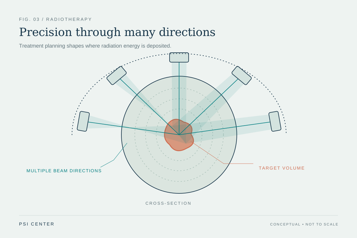
The treatment goal is to deliver the highest prescribed dose to the tumor while sparing nearby normal tissue. Imaging and computer calculations help define the target and shape beams. In three-dimensional conformal therapy, beams from several directions can be arranged to fit the tumor’s outline. Intensity-modulated radiation therapy uses many smaller beams and varies their strength across regions. The physical beam is only one part of a system that includes imaging, geometry and dose planning. [1]
Why electrons fit shallow targets
Suppose a target lies just beneath the skin. A beam that travels deeply may expose more tissue beyond it than necessary. Because electrons have limited range in tissue, they can be suited to some near-surface tumors. The same property becomes a limitation if the target is deep: the beam cannot reach it effectively. The depth of the target, rather than the name of the particle alone, matters to the choice of technique.
This does not mean that electrons are automatically safer or that photons are inherently excessive. Either beam has to be planned for a particular anatomy and treatment purpose. The NCI describes multiple external-beam approaches, each designed to focus prescribed radiation while reducing exposure of normal tissue. The relevant comparison is how a beam’s energy is distributed in the actual treatment geometry, not a simple ranking of particles as better or worse. [1]
Ionizing energy and local treatment
Both photon and electron treatment beams are forms of ionizing radiation: they carry enough energy to damage DNA. That damage is part of how radiation therapy can injure cancer cells, but normal cells in or near the treatment field can also be affected. The National Cancer Institute notes that certain high-energy radiation can damage DNA, while also explaining that risks from medical procedures must be considered alongside their potential benefits. [2]
External-beam treatment does not make a patient radioactive. The radiation comes from a machine and is directed at a local treatment area; once the machine is off, the patient is not emitting the treatment beam. That distinction helps separate radiation entering tissue during treatment from radioactive material remaining in the body. Particle type helps determine where energy can travel, while careful planning shapes where the prescribed dose is delivered. [1]
Explore the physics: an explanatory diagram
Sources & further reading
- External Beam Radiation Therapy National Cancer Institute
- Radiation National Cancer Institute
Prepared for the independent PSI Center reading library. About edition dates and source checking.
This explainer describes scientific principles. It does not assess individual treatment choices.