A Bright Echo Is Not a Diagnosis
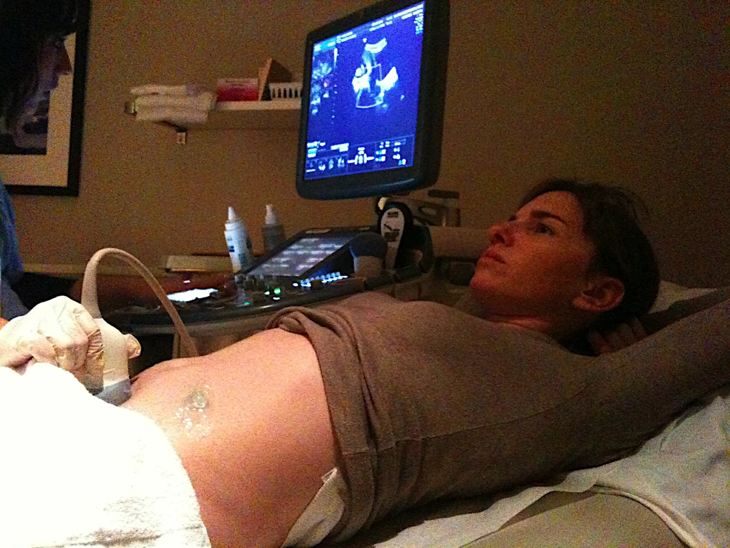
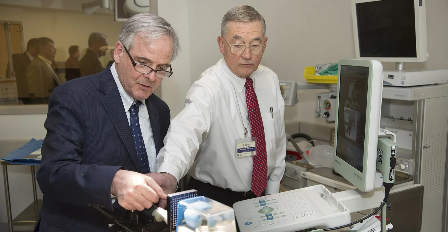
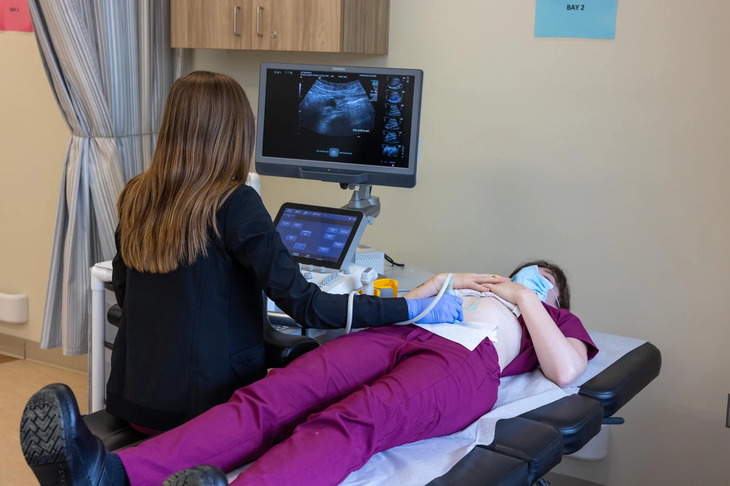
Ultrasound turns returning sound into a useful map, but brightness depends on how waves meet tissue and how scanners display the result. Understanding those steps clarifies both the image’s power and its limits.
A closer look at the science
behind better healthcare.
Follow a question from first principles to the clinic. Browse the science of imaging, treatment, materials, and measurement.
325 articles
Deep dives first, then reading editionsUltrasound turns returning sound into a useful map, but brightness depends on how waves meet tissue and how scanners display the result. Understanding those steps clarifies both the image’s power and its limits.
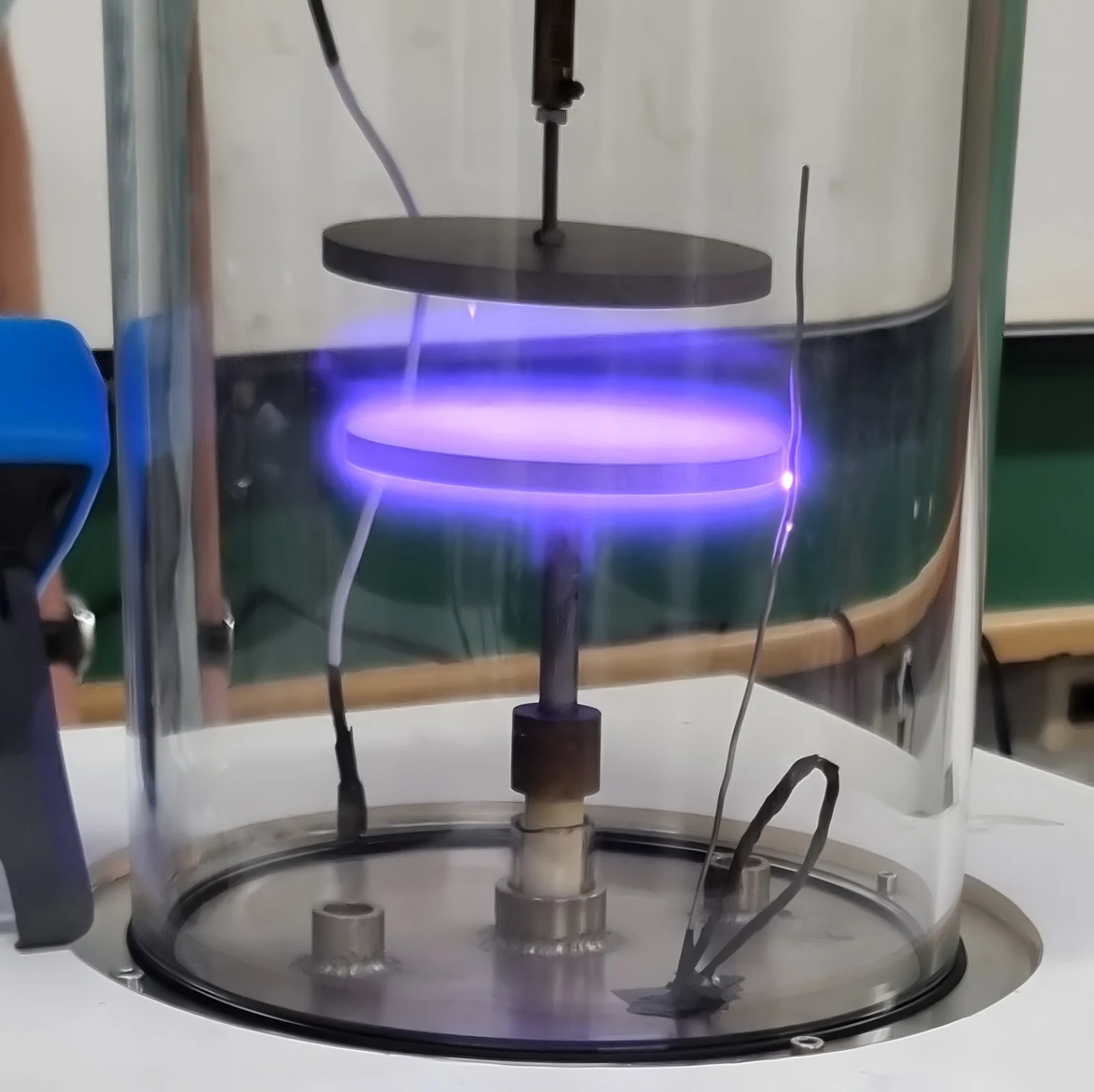
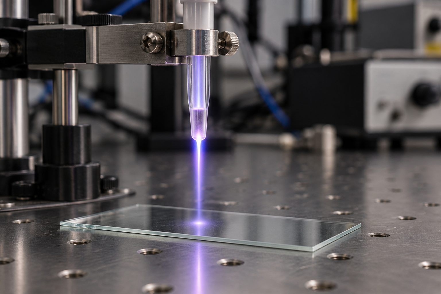
Argon plasma coagulation uses an electrically conducting gas to deliver heat across a small gap. Its surgical role depends on how the gas, electrical field, tissue, and instrument geometry work together.
Near-infrared methods use light absorption and scattering to estimate properties such as hemoglobin concentration and oxygen saturation. Their reach into soft tissue is useful, but signals are indirect and can be affected by skin, motion, and geometry.
Computational models let researchers explore how an imaging device may behave before every question can be answered in a clinic. Their predictions are useful only when grounded in data and checked against physical measurements.
Ultrasound turns echo timing into distance by sending sound pulses and measuring their return. Knowing the speed of sound in tissue lets a scanner place reflecting boundaries at estimated depths.
Plasma is a gas in which some particles carry electric charge. Following what happens to an atom’s electrons explains why plasma responds to fields, conducts current, and appears in both nature and medical equipment.
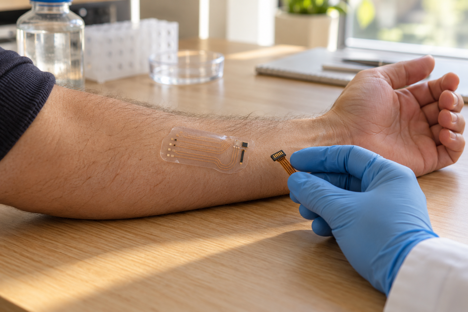
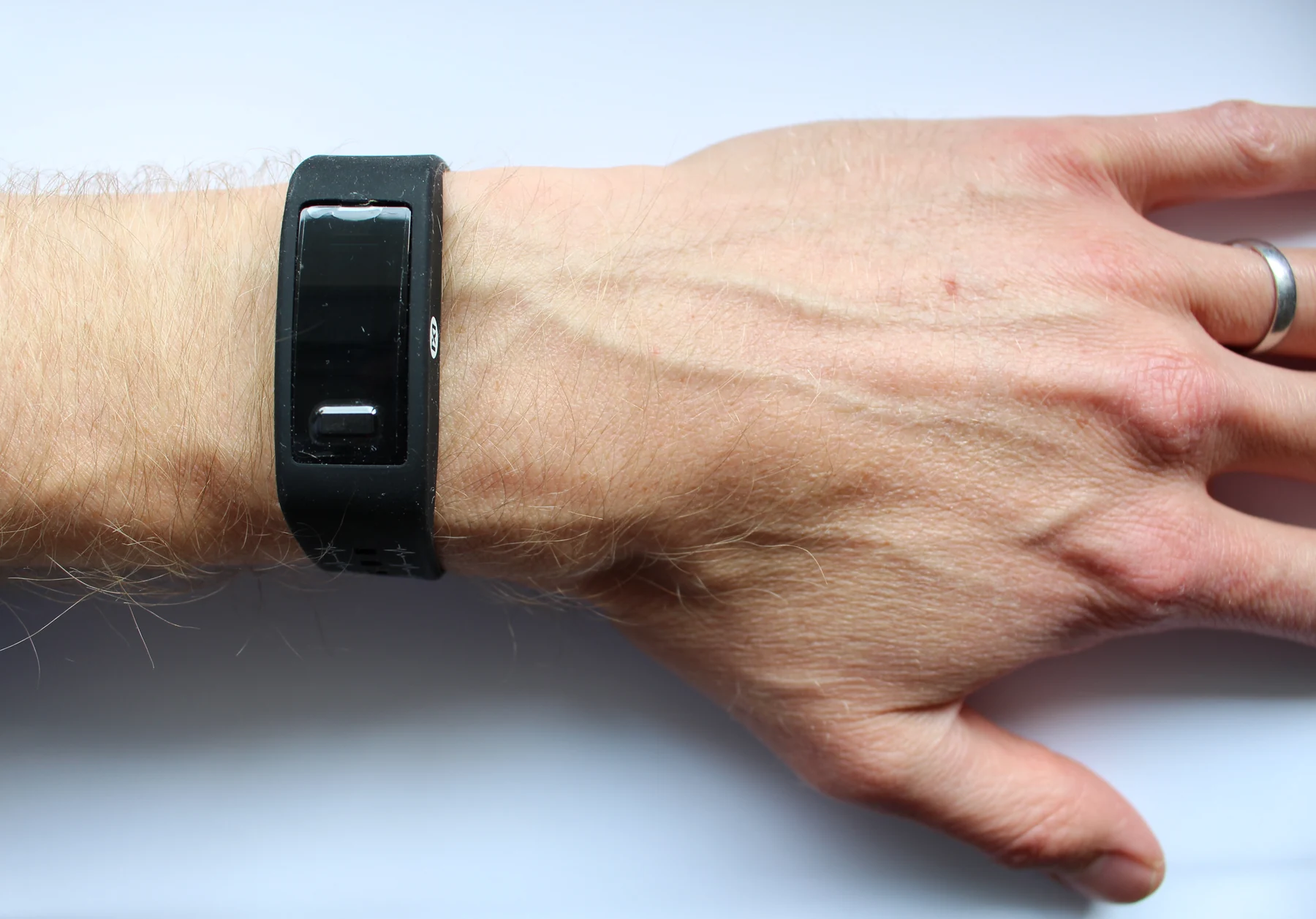
Sensors turn events in the body into signals people can read. Comparing an electrical measurement with a chemical one reveals why the sensing element, conversion step, and interpretation all matter.
A nuclear medicine scan follows a tagged molecule through the body, revealing where biological activity occurs. Its signal can be paired with structural imaging to place that activity in anatomical context.
A sensor reading is a mixture of the quantity of interest and unwanted variation. Filtering can clarify patterns, but the choice of what to suppress affects what a clinician or researcher can still see.
Fluorescence imaging separates a chosen light signal from the surrounding tissue. The method can reveal selected molecules or structures, while the choice of label, tissue depth, and imaging scale shapes what the image can say.
A treatment plan depends on measurements, images, calculations, and patient positioning. Independent checks help catch errors and test whether the planned delivery matches the intended target and constraints.
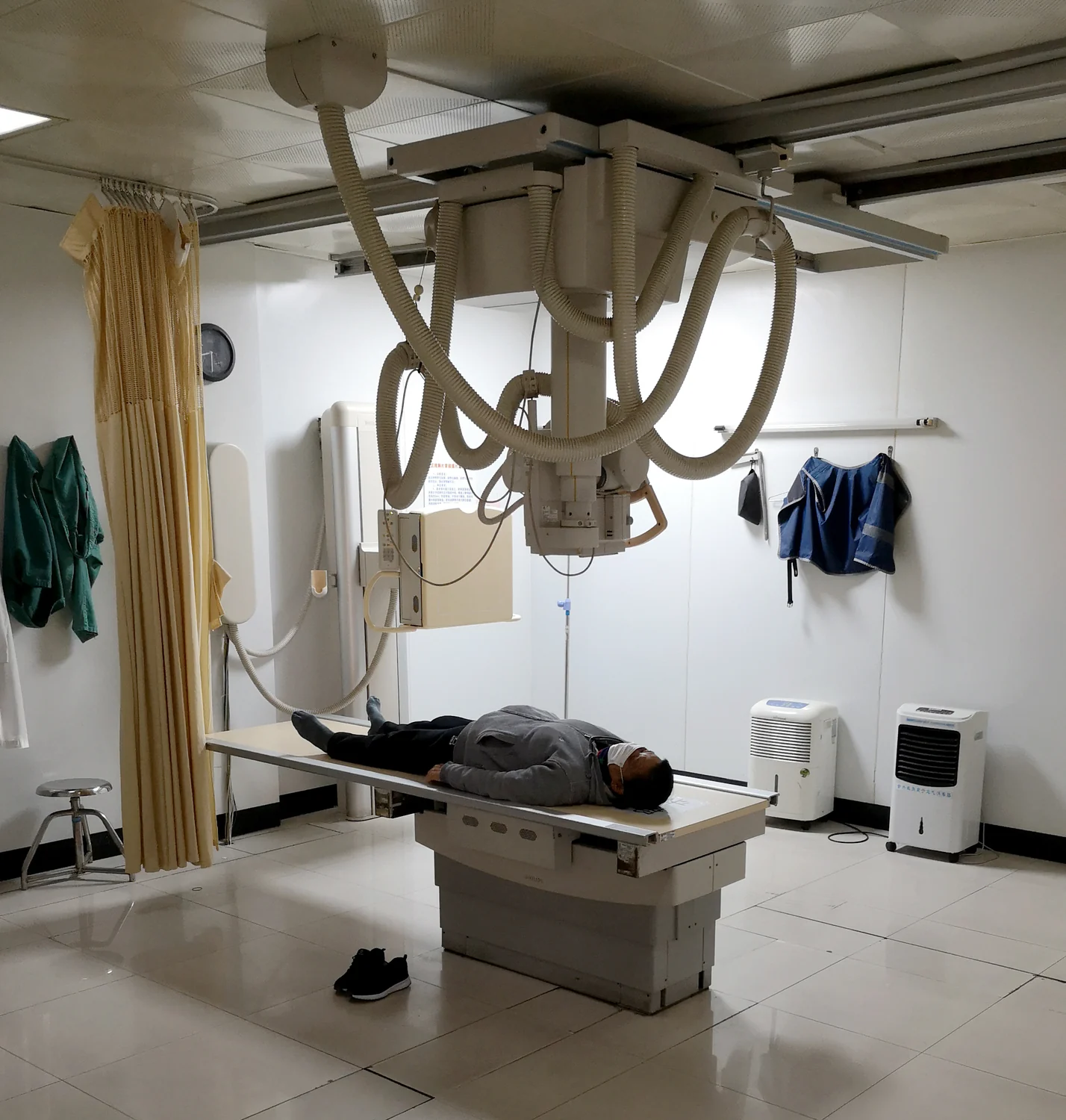
A CT scanner reconstructs cross-sections from many X-ray measurements taken around the body. The changing angles help distinguish overlapping structures that a single projection would collapse into one shadow.
Muscle activity can provide a living control signal for an artificial limb. Sensors and processors translate that activity into movement, while the person’s own muscles remain part of the control loop.
A dialyzer brings blood and dialysis solution close together while a thin membrane keeps them apart. Concentration differences drive some dissolved wastes across, while the machine also manages fluid and mineral balance.
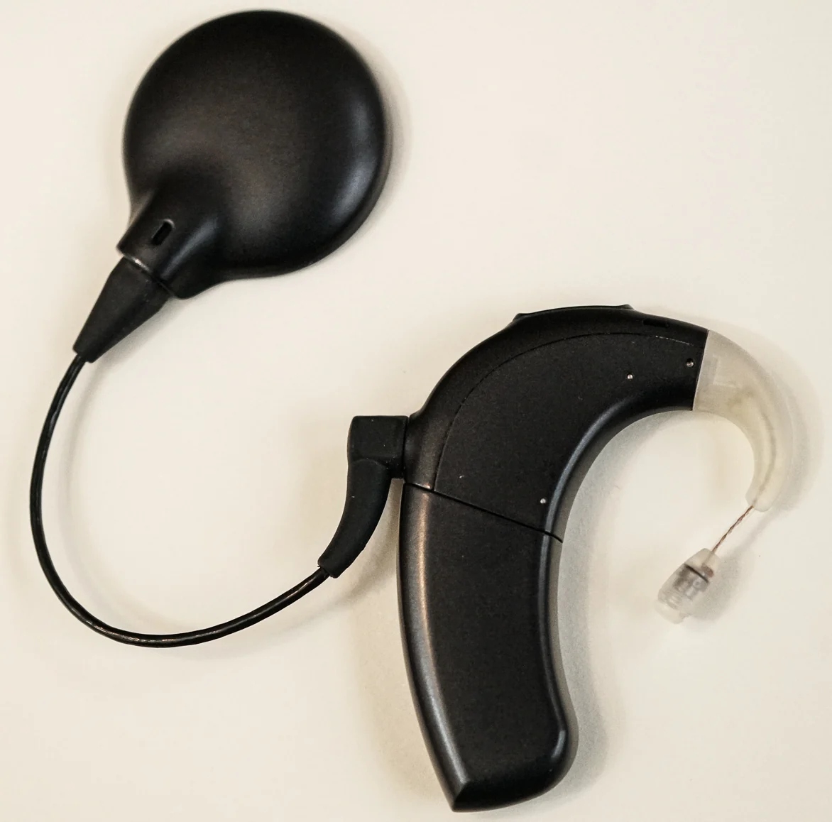
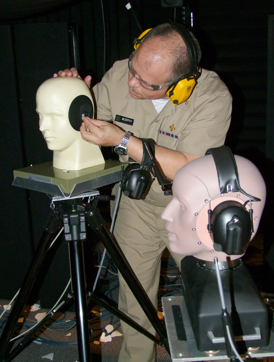
Sound level describes intensity, while exposure duration determines how long the inner ear absorbs that energy. Together with distance and sound character, these factors help explain why a brief impulse and prolonged noise pose different hearing risks.
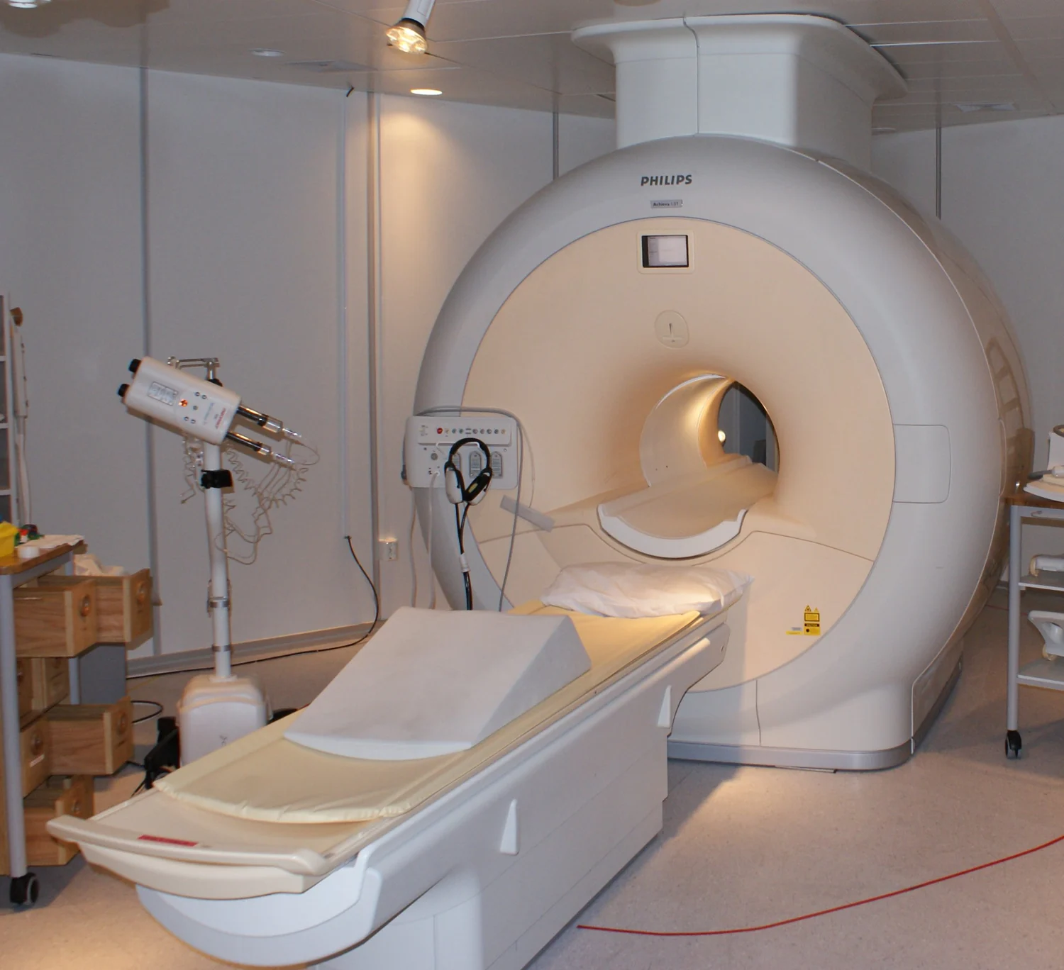
MRI uses a strong magnetic field to organize proton spins and radiofrequency pulses to disturb them. Sensors detect returning signals that vary with tissue chemistry, allowing soft-tissue images without ionizing X-rays.
Systemic radiation therapy uses a carrier molecule to bring radioactive atoms through the bloodstream toward selected cells. Its reach can include many sites, while targeting and nearby-tissue exposure remain central constraints.
A diagram is a compressed account of a physical process. Follow the arrows, identify what is measured, and ask what assumptions connect the drawing to a biological conclusion.