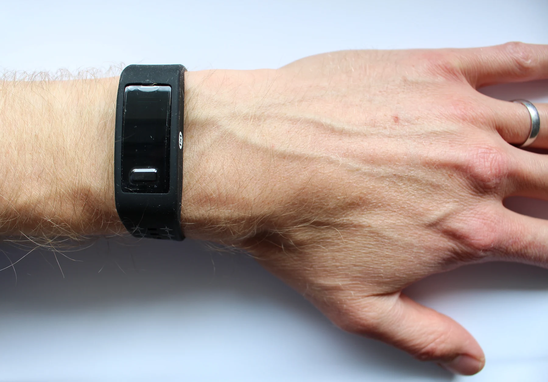
Muscle activity can provide a living control signal for an artificial limb. Sensors and processors translate that activity into movement, while the person’s own muscles remain part of the control loop.
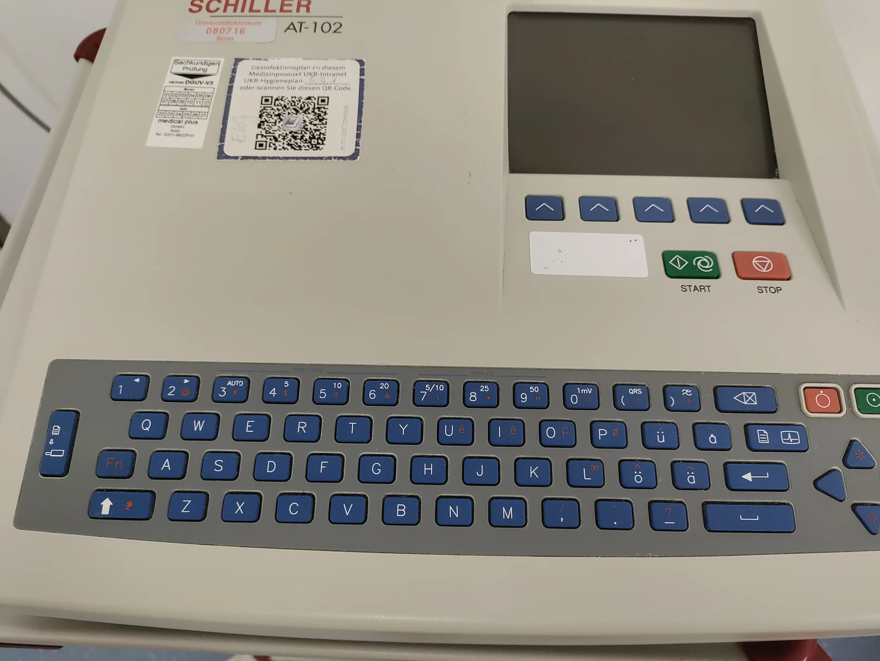
An ECG records voltage differences at the skin, where heart activity is only one contributor. Movement, contact changes and electrical interference can distort the tracing, so signal quality is part of interpretation.
A test detects a target in a particular sample at a particular moment. Its result depends on what the assay measures, how much target is present, and the test’s ability to distinguish signal from absence.
Sensors can remain powered and still become less dependable as their response changes. Calibration, reference chains, and checks against a device’s intended use help reveal whether a long series of readings still means what it once did.
Muscles produce electrical activity as nerves activate them. Surface sensors and needle electrodes sample that activity from different positions, so they answer related but nonidentical questions about nerve and muscle function.
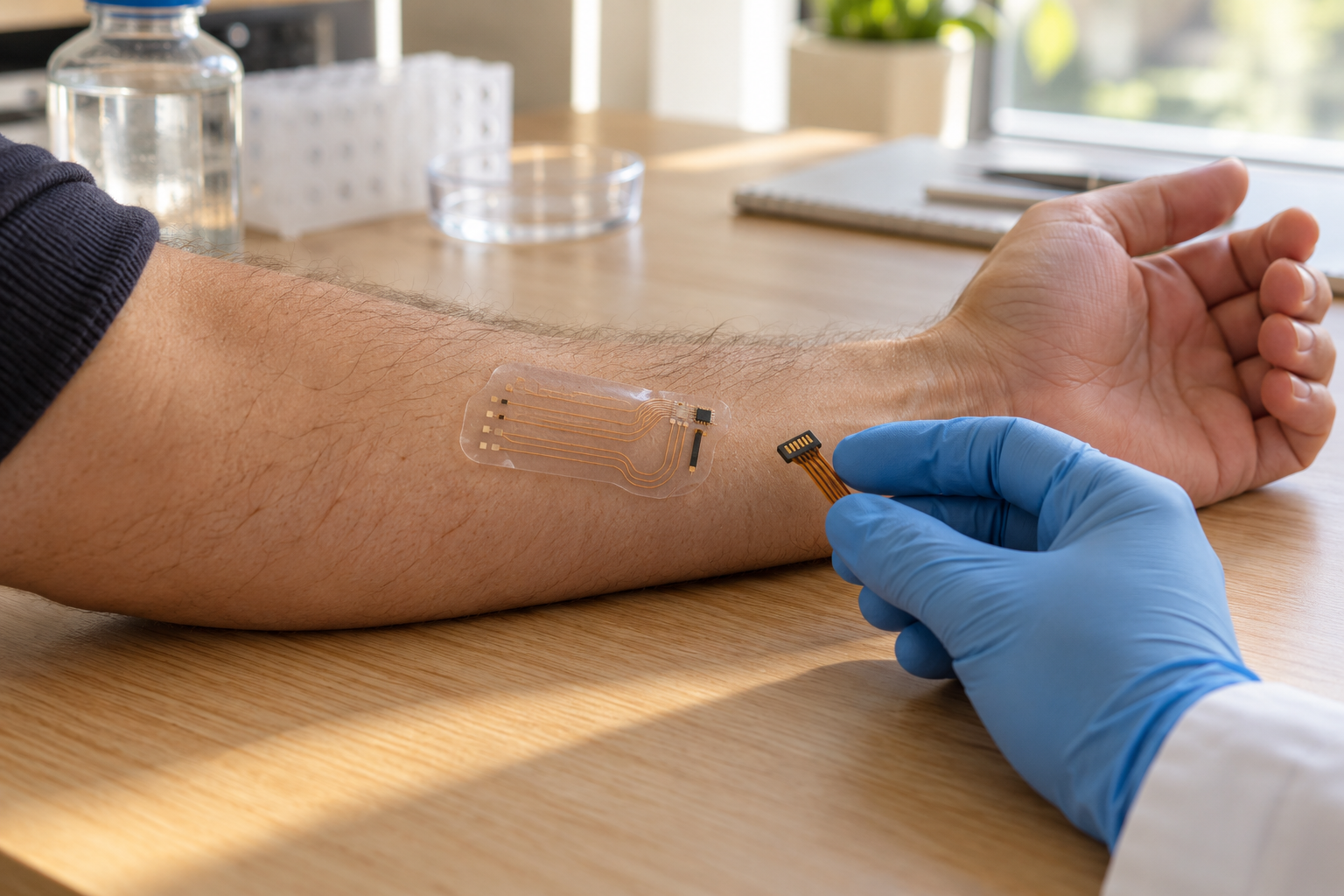
Electrodes record electrical differences at the skin, so contact quality affects how faithfully a small biological signal reaches the instrument. ECG and needle EMG illustrate two distinct ways of sensing activity.
A quick test is the visible end of a longer chain: collecting a sample, converting biological signals into a result, and deciding what that result can support. Understanding each link clarifies both the value and limits of testing near the patient.
Repeated sensor readings can cluster tightly yet sit away from the true value. Separating precision from accuracy clarifies what a device reports and what its pattern of error means for interpretation.
Electromyography records electrical activity in muscle, while nerve conduction testing measures how signals travel along nerves. Together, these measurements can help distinguish patterns of nerve-related and muscle-related weakness.
An ECG electrode records voltage differences at the skin as the heart’s electrical activity spreads. It does not send current into the body, and its tracing is a brief, indirect view of cardiac function.
Microfluidic diagnostics guide tiny fluid volumes through channels where biological chemistry can become a readable signal. Their compactness is useful, but the result still depends on what the test detects and how reliably it distinguishes conditions.
A sensor’s electrical output becomes meaningful only when compared with known references. Calibration builds that bridge, while uncertainty and fitness for purpose determine how much confidence a measurement deserves.
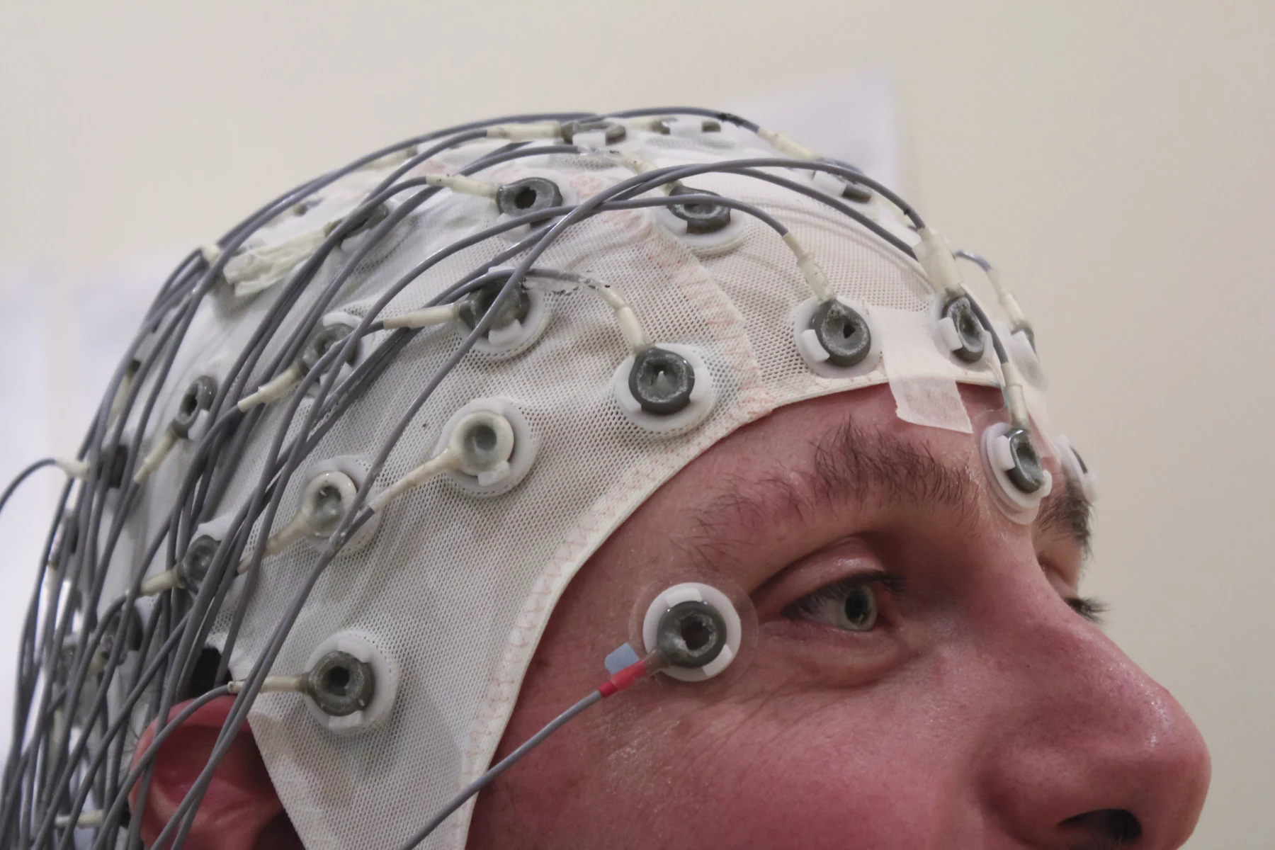
An EEG records voltage patterns at the scalp after signals from many brain cells pass through tissue and meet the outside world. That long path helps explain both the test’s strengths and its sensitivity to movement.
An ECG senses voltage differences at the skin as the heart’s electrical activation spreads and recovers. Its trace reveals timing and rhythm, but a short recording is only a sample of activity, not a diagnosis by itself.
Sensors can collect continuous streams of data, but a health device is useful only when its measurements connect to a meaningful question and a reliable response. Monitoring, diagnosis, and treatment are distinct roles.
Continuous glucose monitors sample fluid between cells, a useful but indirect window on blood glucose. The difference between these compartments helps explain why sensor trends may trail fast changes.
EEG electrodes detect changing voltage patterns at the scalp that reflect coordinated brain activity. The record can support evaluation and monitoring, but it cannot measure intelligence or rule out every seizure.
A sensor’s reading is an inference from physical signals, not a direct window onto the body. Pulse oximetry shows how skin, motion, circulation, and training data can shape errors unequally.