If an ECG line jumps when someone shifts on the bed, did the heart suddenly change? Not necessarily. An electrocardiogram records voltage differences detected by skin electrodes, and those electrodes can also register changes caused by movement, contact or electrical surroundings. These unwanted contributions are called artifacts. Understanding how they enter the recording helps explain why a striking waveform needs interpretation in context rather than automatic attribution to the heart.
The heart creates a changing voltage pattern
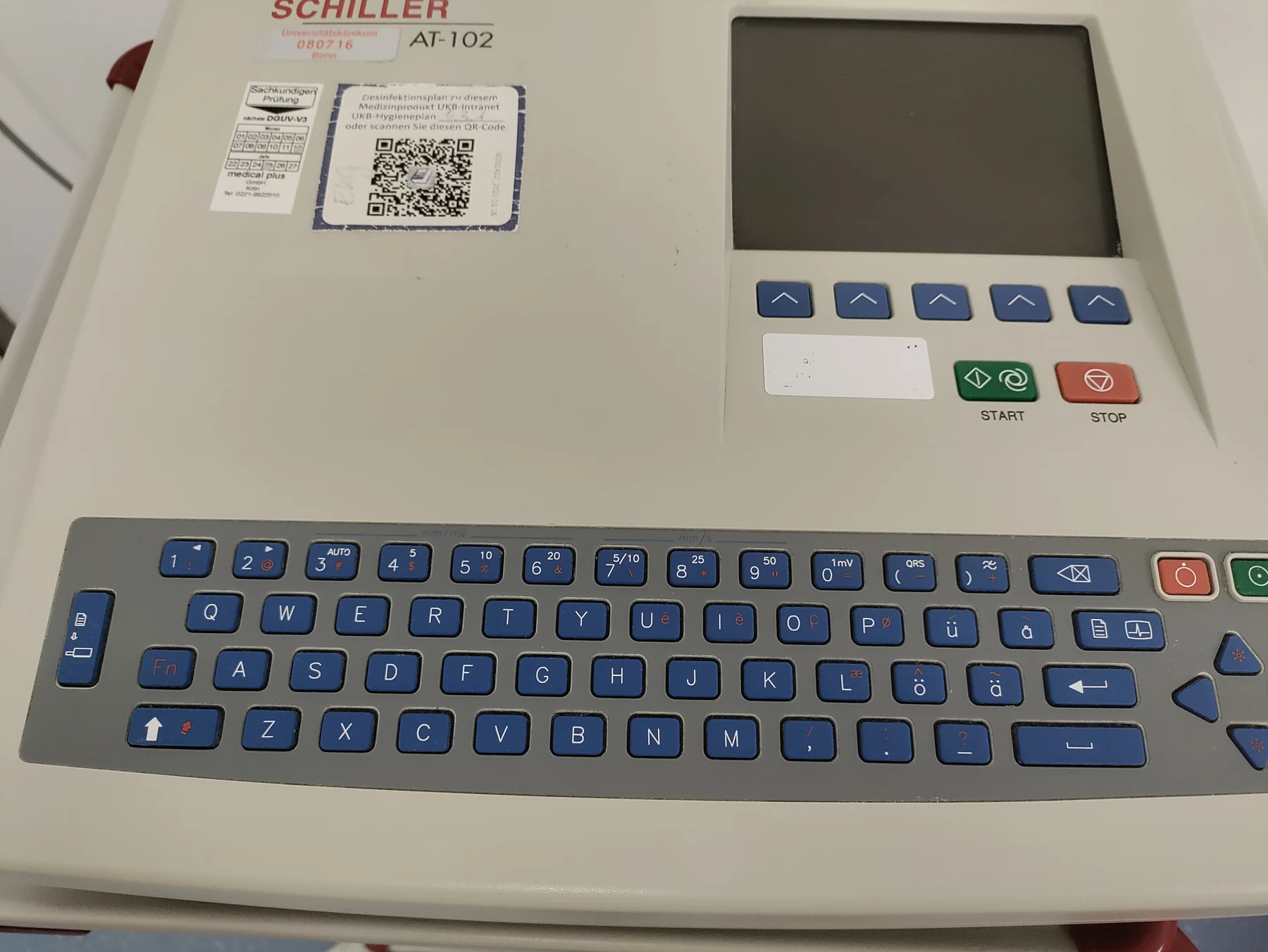
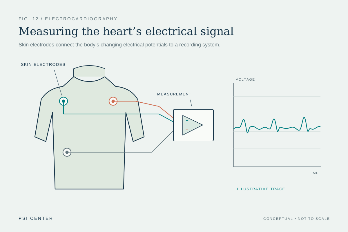
Each heartbeat involves coordinated electrical activity that prompts heart muscle to contract. An ECG, also called an EKG, records the resulting electrical signals through electrodes attached to the skin; it does not send electricity into the body [1]. The familiar tracing includes the P wave, QRS complex and T wave, which correspond to stages of the heart’s electrical cycle. Clinicians use the pattern to assess rate, rhythm and signal timing, among other features [1].
The electrodes do not pick up a single isolated signal from inside the heart. They measure voltage differences between locations on the body, and the machine displays those differences over time. That makes the recording sensitive to the electrical connection between skin and electrode as well as to the heart’s activity. A clean trace is a measurement result shaped by both physiology and the conditions under which it was collected.
Movement changes the measurement
When a person moves, an electrode may shift or the skin beneath it may stretch. That can change contact and alter the voltage the recorder sees, producing a sudden deflection or a noisy baseline. Muscle activity can contribute too: skeletal muscles generate electrical signals when they contract, and those signals may be picked up alongside the heart’s signal. The result can resemble a change in cardiac activity even when the change came from movement or muscle tension.
Imagine recording a quiet conversation with a microphone whose cable is loose. A crackle in the recording might come from the cable rather than the speaker. ECG artifacts are not identical to audio noise, but the analogy captures the central problem: the instrument reports everything reaching its input, not only the source of interest. Recognizing a likely artifact involves looking at its shape, timing, consistency across leads and relationship to movement, while remembering that no one clue settles every case.
Electrical surroundings can add interference
Electrical equipment and wiring can contribute unwanted signals to a recording. This interference may appear as regular oscillations or other patterns superimposed on the ECG. Electrode contact, movement, muscle activity and external electrical noise can overlap, so a distorted tracing may have more than one cause. The practical goal is to distinguish the heart’s organized pattern from disturbances introduced by the body–electrode interface or surroundings.
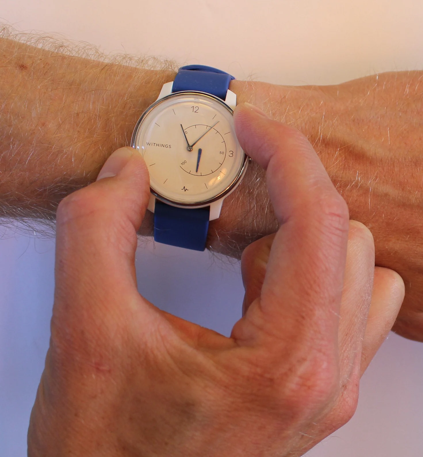
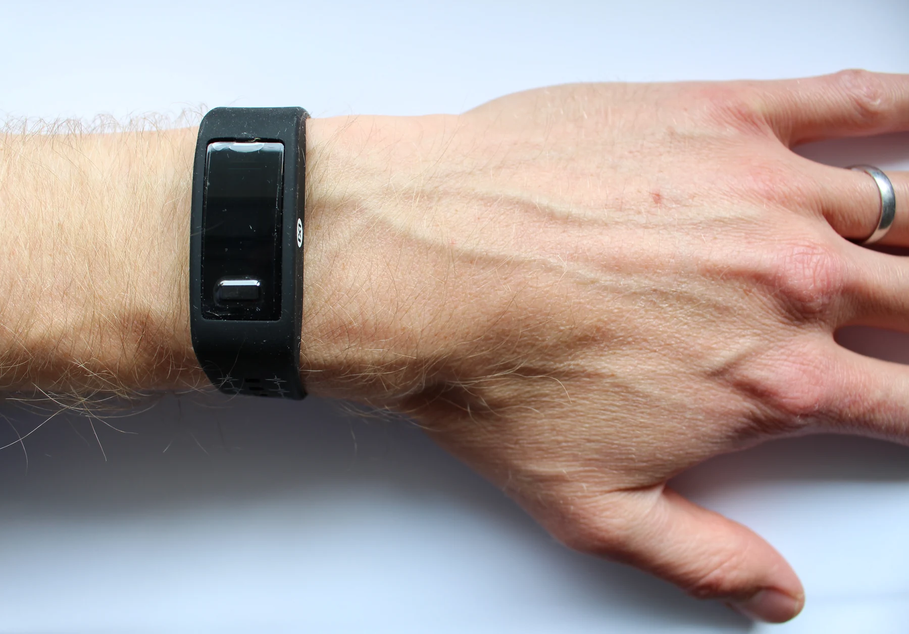
A digital recorder samples the changing voltage and stores or displays it as data. Digital processing can help filter certain kinds of noise, but filtering is not a magic eraser: a true cardiac feature may share frequencies or shapes with an artifact. The NIBIB overview of digital health describes how sensors and connected systems collect and transmit health information, including wearable monitoring examples [2]. Those systems can expand monitoring beyond a clinic, but collected data still depend on sensor placement, signal quality and interpretation.
A tracing is evidence, not a diagnosis by itself
An ECG is a short recording when performed as a standard test. If symptoms come and go, longer monitoring may capture activity missed in that snapshot; Holter and event monitors are examples of ways to record for longer periods [1]. More recording time may reveal intermittent patterns, but it also means more opportunities for motion and environmental noise. The signal needs to be read with its quality and circumstances in mind.
Digital tools can flag patterns or help organize data, but the presence of a sensor or algorithm does not make every wearable a diagnostic device. NIBIB describes wearables as having potential roles in monitoring and self-management, alongside specific research examples and developing technologies [2]. An ECG remains a measurement of electrical signals at the body surface, and its interpretation may be considered with symptoms, history and other tests [1]. Separating cardiac activity from artifact is therefore part of the science of measurement, not an optional cleanup step.
Explore the physics: an explanatory diagram
Sources & further reading
- Electrocardiogram MedlinePlus
- Digital Health NIBIB
Prepared for the independent PSI Center reading library. About edition dates and source checking.
This explainer describes scientific principles. It does not assess individual treatment choices.