What does a growing tissue need from a material that is meant to disappear? Often, it needs a temporary place to attach, organize, and exchange signals with neighboring cells. In tissue engineering, that supporting material is called a scaffold. It can be imagined as architecture at a scale far below what the eye can see: pores, fibers, and surfaces give cells a local environment while researchers investigate how living tissue might form around or within them.
A framework at cell scale
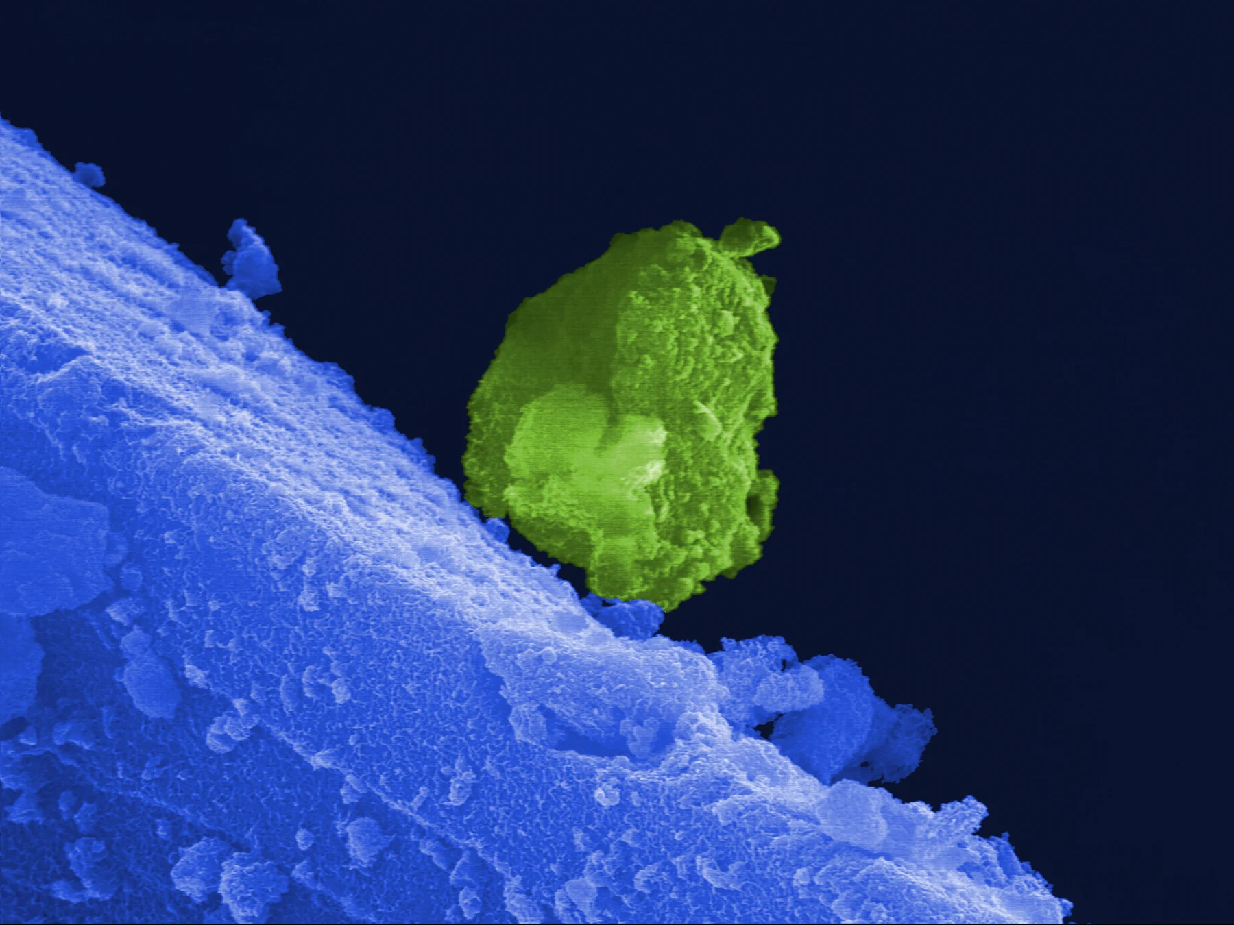
Many scaffolds imitate aspects of the extracellular matrix, or ECM: the network of proteins and other molecules surrounding cells in living tissue. Cells do not experience this network as a passive container. Its physical structure and chemistry help shape the environment in which cells attach and interact. Biomaterials can be natural or synthetic and can be formed as fibers, foams, films, or other structures for biomedical use [1].
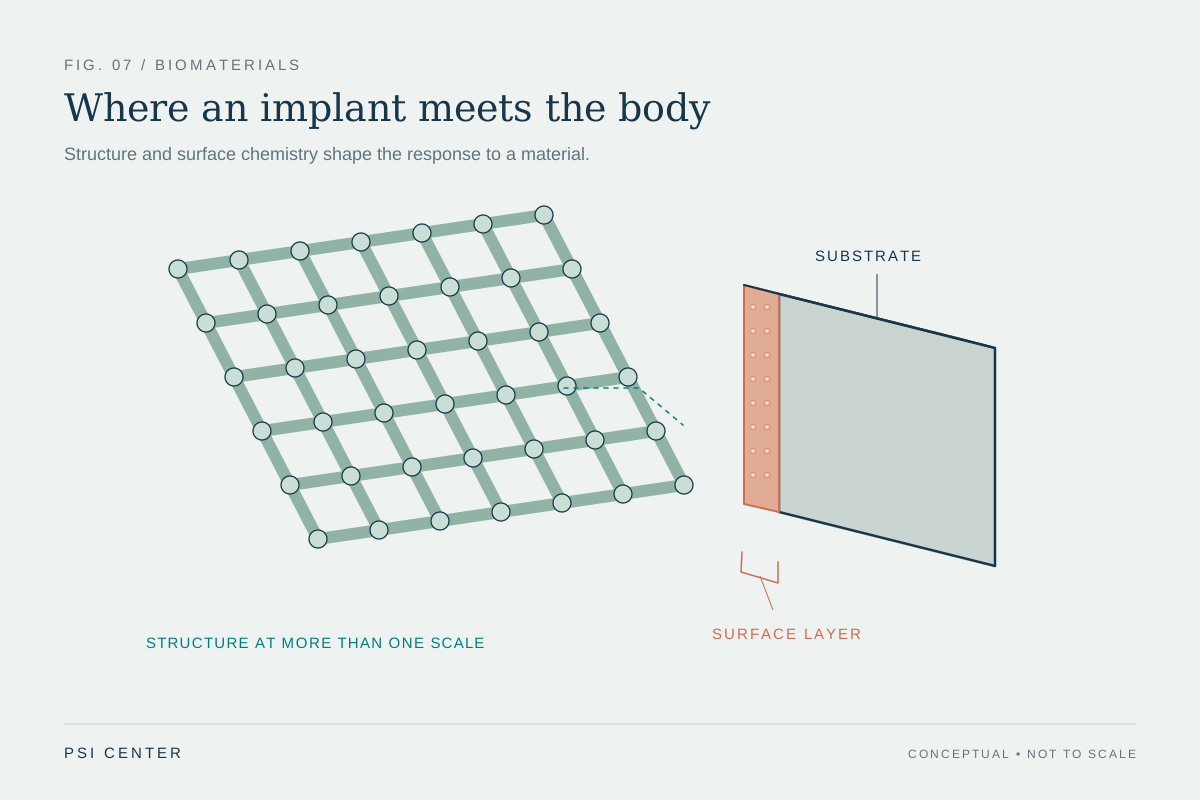
A scaffold’s geometry matters because cells need surfaces and spaces to occupy. A web of fibers, for instance, offers a different pattern of attachment from a solid sheet. The exact design depends on the intended tissue and the material. Calling a scaffold temporary architecture is useful because it emphasizes both its support and its limits: the material helps organize a process, but it is not itself the finished tissue.
Cells read mechanical and chemical cues
Cells encounter a material through more than its shape. Stiffness, surface chemistry, and composition can affect how a cell attaches and what local signals it receives. Biomaterials designed to match features of native ECM may encourage tissue integration, while a mismatch can contribute to the body’s foreign body response—a reaction in which the immune system attempts to degrade or isolate an implanted material [1].
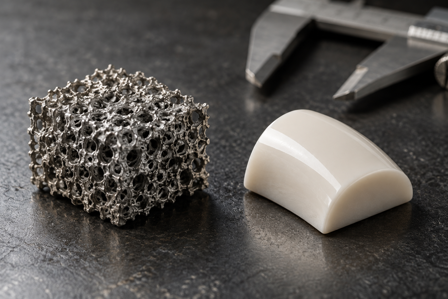
Consider two imagined scaffolds with the same outer dimensions. One has a porous fibrous interior; the other is dense and smooth. Cells could contact and organize within the first in ways that the second does not allow. This is a mental model, not a guarantee that one design will succeed clinically. The actual outcome also depends on the cells, the target tissue, the material’s behavior in the body, and how the construct is made.
A scaffold must be measured as well as designed
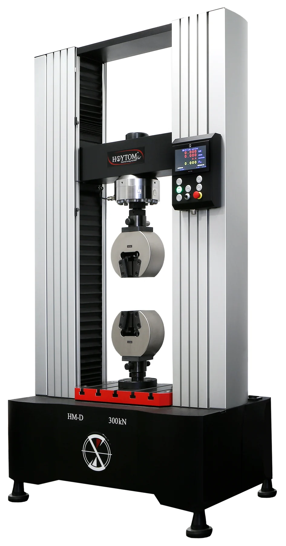
A promising structure is not enough if its properties vary unpredictably from batch to batch. Tissue engineering therefore depends on reliable ways to characterize cells, biomaterials, scaffolds, and the tissue-engineered constructs built from them. NIST describes its work as developing tools and standards for measuring such properties, because consistency and quality matter as regenerative medicine moves toward practical manufacturing [2].
Some questions are very concrete: How wide are the scaffold’s fibers? Are cells inside a hydrogel scaffold still viable? What does a three-dimensional image reveal about how cells and scaffold interact? NIST identifies projects addressing nanofiber measurement, cell viability in hydrogels, and three-dimensional cell–scaffold interactions [2]. These measurements help researchers compare designs and identify variation; they do not by themselves establish that a material will repair tissue in a patient.
From laboratory construct to living repair
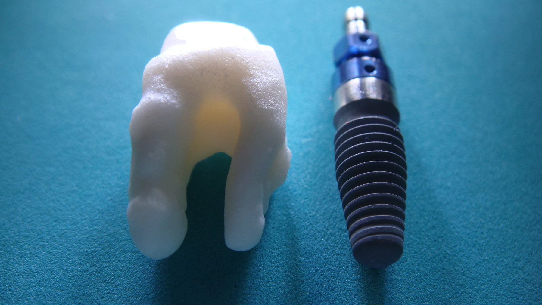
The scaffold is one part of a larger system. Cells must survive, interact with the material, and form tissue with useful properties. The body may also respond to an implanted material in ways that change its performance over time. Materials science, cell biology, and manufacturing therefore meet at the scaffold’s surface and within its pores. Biomaterials already serve in varied devices, but a tissue-engineered scaffold intended to help restore damaged tissue remains a distinct kind of application [1].
Research examples show both the promise and the careful steps involved. A biodegradable polymer film designed to produce electrical current under pressure was studied with exercise in a rabbit model of osteoarthritis; the reported tissue regeneration in that model does not establish a proven treatment for people [1]. The path from temporary framework to dependable clinical product requires reproducible materials, meaningful tests, and evidence that the resulting tissue works as intended.
Explore the physics: an explanatory diagram
Sources & further reading
- Biomaterial Technologies NIBIB
- Tissue Engineering NIST
Prepared for the independent PSI Center reading library. About edition dates and source checking.
This explainer describes scientific principles. It does not assess individual treatment choices.