How can sunlight alter skin when ultraviolet light is invisible? The answer begins with photons: packets of electromagnetic energy that carry different amounts depending on their wavelength. UV sits between visible light and X-rays on the electromagnetic spectrum, but it does not behave as one uniform beam. Its wavelength affects how far it penetrates, and its energy can disturb molecules in skin cells.
Energy arrives in packets
Electromagnetic radiation travels as waves and can also be described as photons. A wave’s wavelength is the distance between successive peaks. Shorter wavelength UV photons carry more energy than longer wavelength ones, though UV remains distinct from the higher-energy X-rays in the spectrum. This difference helps explain why UV has biological effects that visible light does not share in the same way. [1]
The word radiation can sound as if all forms pose the same hazard. It does not. Ionizing radiation, including X-rays and gamma rays, has enough energy to damage DNA directly; lower-energy, non-ionizing radiation is a different category. UV is non-ionizing, yet UVA and UVB can still damage skin through their interactions with tissue molecules. [2]
Wavelength changes the route
Imagine two narrow beams reaching the same patch of skin. UVB has the shorter wavelength and primarily reaches the epidermis, the outer skin layer. UVA has a longer wavelength and can penetrate into the dermis beneath it. The distinction is about where energy is deposited, not a guarantee that one band is harmless: both UVA and UVB can damage skin. [1]
The sunlight reaching people is mostly UVA with a smaller amount of UVB because the atmosphere absorbs all solar UVC and some UVB. UVC, the most energetic portion of UV, does not reach Earth’s surface from the sun; artificial lamps and lasers can produce it. Certain UVC exposures can injure eyes and burn skin, while some lamps also emit UVB or generate ozone, so their hazards are not captured by the label “UV” alone. [1]
From exposure to biological change
A useful mental model is to picture a molecule as a small structure held together by particular chemical bonds. When UV energy is absorbed, it can change those structures and interfere with normal cellular processes. The visible redness of sunburn signals short-term overexposure; repeated or prolonged UV exposure can contribute to premature skin aging and skin cancer. Those outcomes reflect biological damage accumulated through exposure rather than a beam simply heating the whole body. [1]
The amount reaching skin varies with conditions. UV is stronger at higher altitudes and near the equator, can remain present under clouds, and can reflect from snow, sand, pavement, and water. Time of day and season also matter because the sun’s angle changes how much atmosphere the rays cross. The UV Index summarizes expected skin-damaging UV at a location, helping translate an invisible spectrum into a practical estimate of intensity. [1]
A dose can be useful and risky
UV exposure is not biologically one-sided. UVB helps skin produce vitamin D3, which contributes with calcium to bone and muscle health, but the exposure associated with that benefit varies with factors such as skin color, diet, clothing, latitude, and season. A simple “more is better” rule would ignore the same capacity of UV to injure skin. [1]
Phototherapy uses carefully monitored UV exposure for selected conditions, including some severe cases of psoriasis, eczema, and vitiligo that have not responded to other approaches. It is supervised by health professionals, and the dose is managed because treatment does not erase UV’s side effects. The physical principle is the same interaction of photons and tissue; the difference is controlled use, with a specific clinical aim and attention to risk. [1]
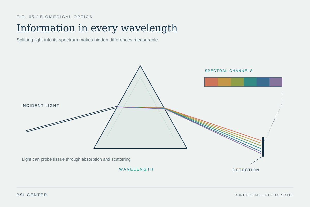
Explore the physics: an explanatory diagram
Sources & further reading
- Ultraviolet Radiation FDA
- Radiation National Cancer Institute
Prepared for the independent PSI Center reading library. About edition dates and source checking.
This explainer describes scientific principles. It does not assess individual treatment choices.