What makes ultraviolet light different from red light if both travel through empty space at the same speed? The answer lies in the spacing of their waves and the energy carried by each photon. A photon is a discrete packet of electromagnetic energy; wavelength measures the distance between repeating wave peaks. Shorter wavelength means higher photon energy, a pattern that helps organize the spectrum from radio waves through visible light to X-rays. [1]
Waves have a spacing
Picture a row of evenly spaced crests moving across water. The distance from one crest to the next is the wavelength. Electromagnetic waves—including radio, visible light, ultraviolet, and X-rays—also have wavelengths, though they do not need water or air to travel. They are made of photons that move at the speed of light and behave in wave-like ways. [1]
Color gives a familiar visible example. Red light has a longer wavelength than violet light. Beyond violet lies ultraviolet, with shorter wavelengths; beyond ultraviolet lies X-ray radiation, shorter still. The electromagnetic spectrum is a continuous range, even though we divide it into named bands for convenience. The boundaries help describe how radiation behaves, but they do not create a gap in nature.
Shorter waves carry more energy per photon
Photon energy is inversely related to wavelength: as wavelength gets shorter, the energy of each photon rises. This can be expressed with the relationship E = hc/λ, where E is energy, h is Planck’s constant, c is the speed of light, and λ is wavelength. A reader need not calculate values to use the idea: a shorter wavelength means more energy in each photon. The FDA describes the spectrum in the same terms, linking short wavelengths to more energetic photons. [1]
That does not mean a beam’s total energy depends only on wavelength. The number of photons also matters. A dim beam can contain fewer photons than a bright one, even if each photon in the dim beam carries more energy. This distinction between energy per photon and the total energy delivered is useful whenever brightness and wavelength are being compared.
Why the distinction matters in tissue
Visible light and ultraviolet are both non-ionizing radiation, while X-rays and gamma rays are ionizing. Ionizing radiation has enough energy to remove electrons from atoms or molecules; the NCI explains that X-rays and gamma rays can directly damage DNA or cells. Non-ionizing radiation includes visible light and ultraviolet, though UV can still harm tissue through its interactions with molecules. [2]
For example, the FDA notes that UVA and UVB can damage skin, with sunburn a sign of short-term overexposure and prolonged exposure associated with premature aging and skin cancer. UVC photons have higher energy than UVA photons because UVC has shorter wavelengths, but UVC from the sun is blocked by the ozone layer. Artificial UVC sources can injure skin and eyes. Wavelength helps predict interaction, but exposure and biological context also matter. [1]
A spectrum is a map, not a safety scale
It is tempting to rank electromagnetic waves as harmless at one end and dangerous at the other. Energy per photon is an important physical difference, but the effect on a person depends on how much radiation reaches tissue, its duration, wavelength, and the tissue’s response. The FDA notes that supervised UV phototherapy uses carefully monitored doses for certain conditions, while UV overexposure can be harmful. The same wavelength category can therefore appear in very different contexts. [1]
The spectrum is best thought of as a map connecting wave spacing to photon energy. Long radio wavelengths correspond to low energy per photon; visible light sits between infrared and ultraviolet; X-rays have shorter wavelengths and more energetic photons. That map helps explain why different bands are used for communication, illumination, imaging, or treatment, while also reminding us that the label alone does not describe the dose or outcome. [2]
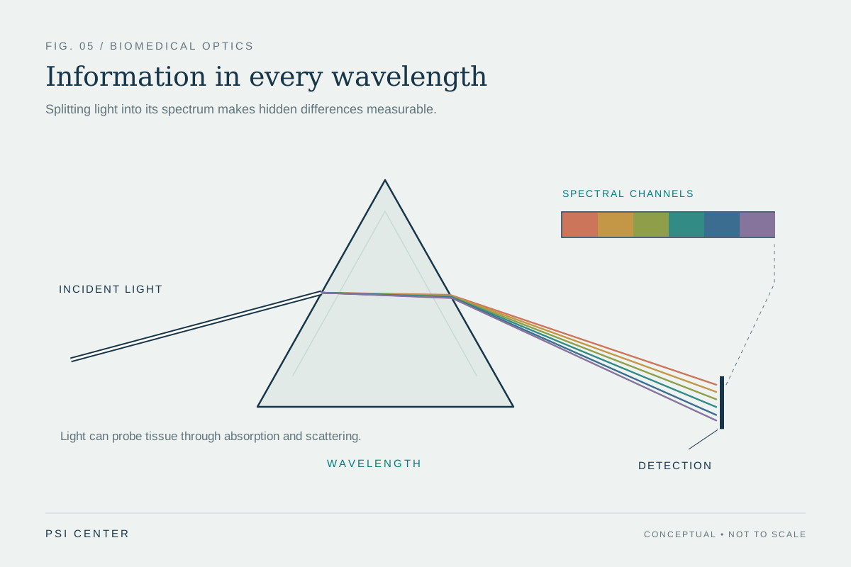
Explore the physics: an explanatory diagram
Sources & further reading
- Ultraviolet Radiation FDA
- Electromagnetic Fields and Cancer National Cancer Institute
Prepared for the independent PSI Center reading library. About edition dates and source checking.
This explainer describes scientific principles. It does not assess individual treatment choices.