How can a beam reach a tumor and then stop before traveling farther through the body? Proton therapy uses positively charged particles that lose energy as they pass through tissue, with a finite range. That feature can concentrate dose near a target, but it also makes the depth at which protons stop a crucial planning question. The beam must be aimed in a body whose tissues and position shape its path.
A beam with a stopping point
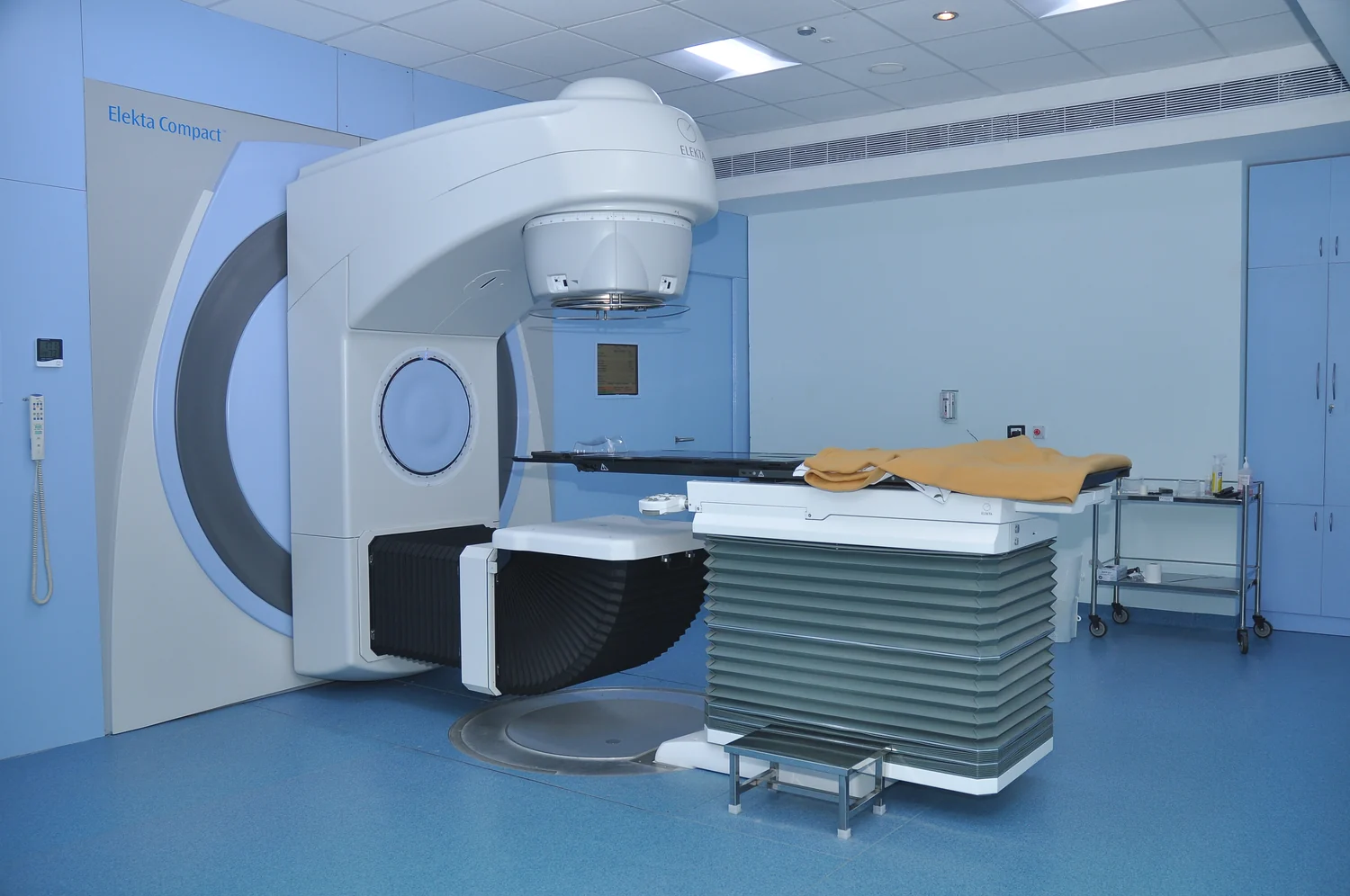
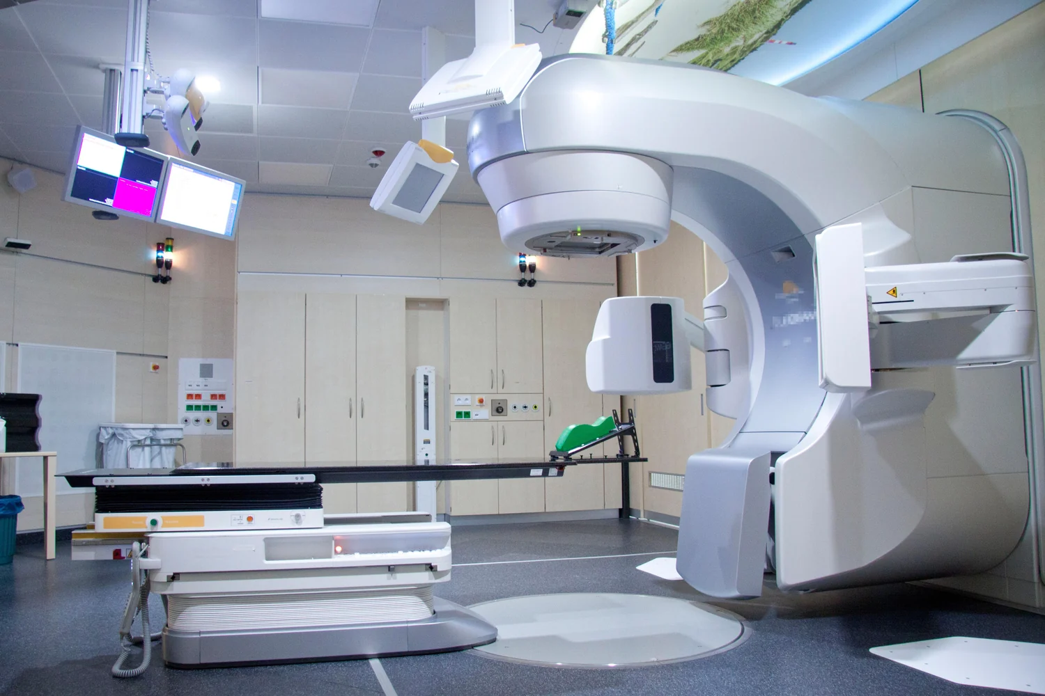
External beam radiation therapy directs radiation from a machine at a defined region of the body. Photon beams, used in many radiation machines, can reach deep tumors but scatter some radiation along their path and continue beyond the tumor into normal tissue. Proton beams also reach deep targets, but they can stop once they reach the tumor. This difference is why clinicians consider whether protons might reduce radiation exposure to surrounding normal tissue. [1]
A useful mental picture is to imagine two routes through a crowded room. A photon beam leaves some energy along its whole route and continues past the target; a proton beam can release much of its energy near the intended endpoint. But the endpoint is not a painted mark in the body. It depends on the material the particles traverse. Bone, soft tissue, and air-filled spaces do not present identical paths, so the planned range must be tied to the patient’s anatomy.
Images become a map for the beam
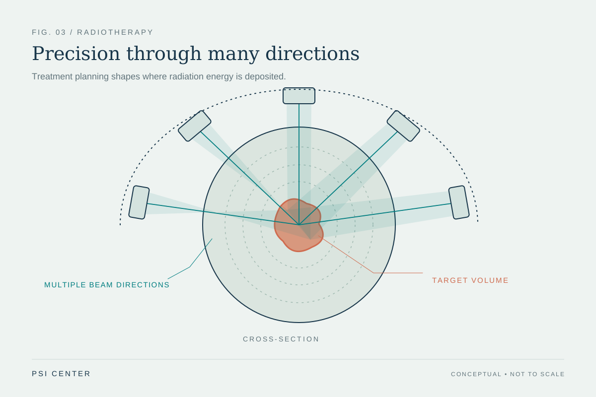
Before treatment, imaging helps define where the tumor is and what lies around it. In three-dimensional conformal therapy, scans such as CT, MRI, or PET are used to plan the treatment region; a computer designs beams from multiple directions to fit the target’s shape. Other approaches divide the beam into many smaller parts and vary their strength. The central task is to deliver the prescribed dose to the tumor while sparing nearby normal tissue. [1]
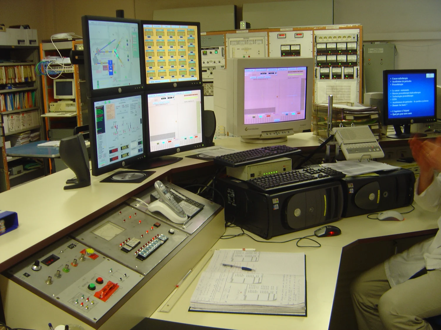
Computational modeling is a way to simulate a complex system using mathematics, physics, and computer science. A treatment model can calculate predicted dose patterns under different conditions, using a representation of anatomy and the beam. Such a prediction is useful because it allows planners to compare possibilities in a virtual setting. It remains a model of reality, however: the quality of its output depends on its inputs and on how well the model represents the physical situation. [2]
Position and motion alter the path
Even a carefully drawn target can shift relative to the beam. The body’s posture must be reproduced from planning through treatment, which is why positioning marks, masks, or molds may help hold a person in the same arrangement. Some tumors move with breathing or digestion. Radiation techniques may use repeated imaging to detect changes in a tumor’s size or location and allow position or dose adjustments when needed. Motion matters because a small shift can change where a finite-range beam reaches its endpoint. [1]
Consider a proton path planned through tissue to a tumor near a sensitive structure. If the person’s position or the anatomy differs from the planning image, the beam encounters a changed route. The physical range does not know where the tumor was on yesterday’s scan; it responds to the material in front of it now. This is why treatment planning and image guidance are connected parts of controlling where energy is deposited.
Precision is a goal, not a guarantee
Protons’ stopping behavior offers a physical advantage to investigate, but it does not mean proton therapy is automatically safer or better for every tumor. The National Cancer Institute notes that clinical trials are comparing proton and photon approaches, and that proton equipment is large and costly. Treatment selection depends on the cancer, its location, and the treatment plan rather than a single appealing property of the beam. [1]
The planning problem is therefore a balance: define the target from images, model how radiation travels through tissue, reproduce the patient’s position, and account for anatomy that moves or changes. Models can help explore many conditions and focus attention on important experiments or decisions, but they do not erase uncertainty. The proton’s range is valuable precisely because it is finite; that same finiteness makes careful planning essential. [2]
Explore the physics: an explanatory diagram
Sources & further reading
- External Beam Radiation Therapy National Cancer Institute
- Computational Modeling NIBIB
Prepared for the independent PSI Center reading library. About edition dates and source checking.
This explainer describes scientific principles. It does not assess individual treatment choices.