Why can one ultrasound system help clean a surgical instrument while another produces an image of a heart? Both use sound waves above the range of human hearing, but the resemblance ends at the physics of vibration. A cleaning bath uses acoustic energy in liquid to loosen material from an object. Medical imaging sends sound into the body and interprets returning echoes as information about internal structure and motion.
Sound waves carry energy
Sound is a mechanical disturbance: pressure changes move through a material such as air, water, or tissue. Ultrasound simply means sound at frequencies higher than people can hear. Its effects depend on frequency, intensity, pulse pattern, the surrounding material, and the object it encounters. A wave in water can shake loose debris; a wave that returns from a tissue boundary can help locate that boundary.
The word “ultrasound” therefore names a range of sound, not a single medical function. A bath designed for instrument cleaning and a probe used for imaging have different transducers, geometry, settings, and intended outcomes. Treating them as interchangeable because both vibrate at ultrasonic frequencies would be like assuming a microphone and a loudspeaker do the same job because each handles sound.
How an ultrasonic cleaner loosens soil
In an ultrasonic cleaner, acoustic waves pass through a liquid. Their pressure changes can create and collapse tiny bubbles, a process called cavitation. The resulting agitation disrupts the bonds holding particulate soil to a surface, helping fluid reach grooves and small spaces [1]. The bath removes contaminants from an object; it does not create an image of the object's interior.
Cleaning is one step in reprocessing, not the same as disinfection or sterilization. The CDC notes that thorough cleaning is needed before high-level disinfection or sterilization because organic and inorganic residue can interfere with those later processes [1]. A cleaner's performance also depends on whether liquid contacts the relevant surfaces. Hinges may need to be open and instruments arranged so solution can reach them.
How diagnostic ultrasound makes an image
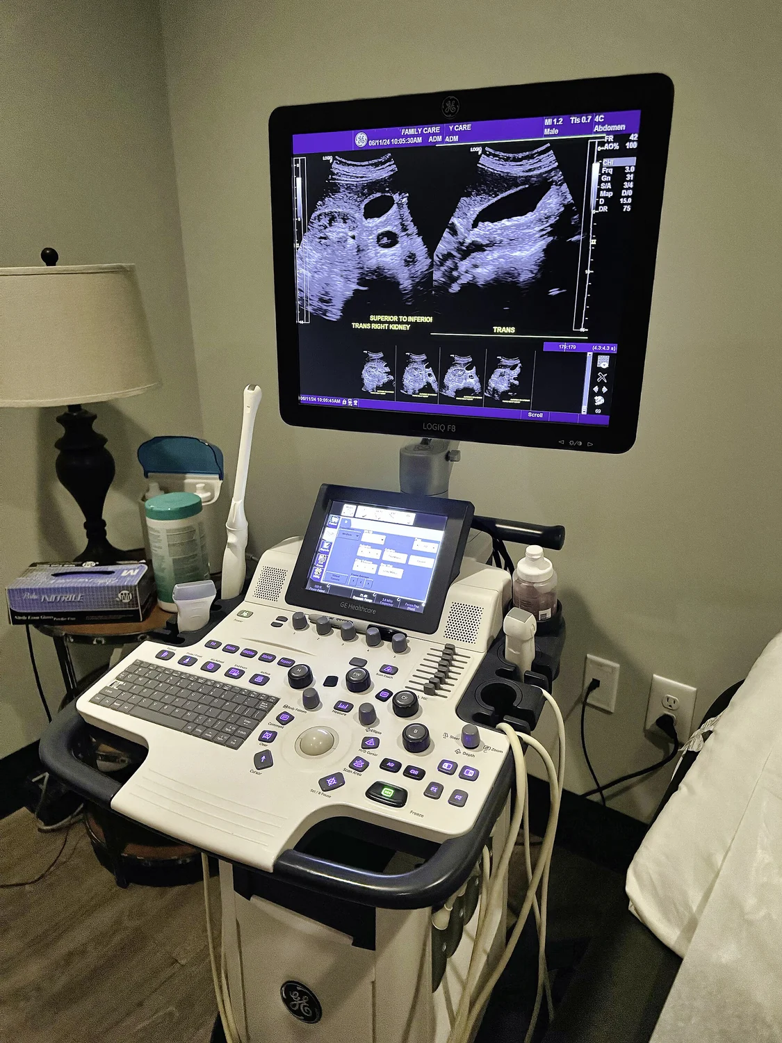
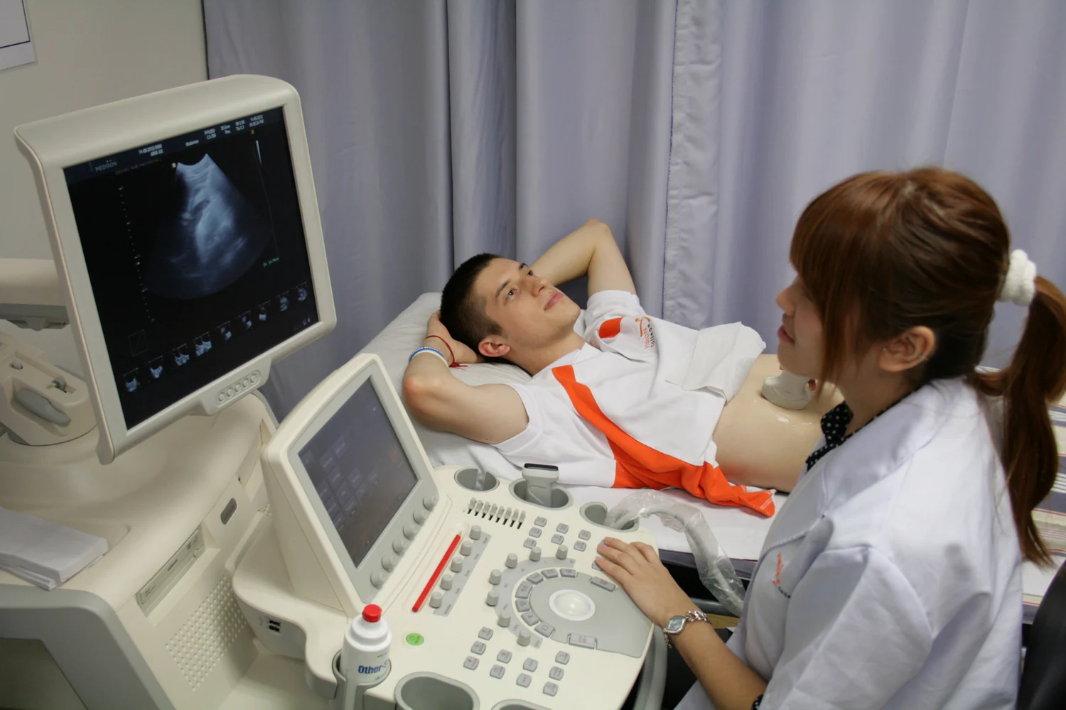
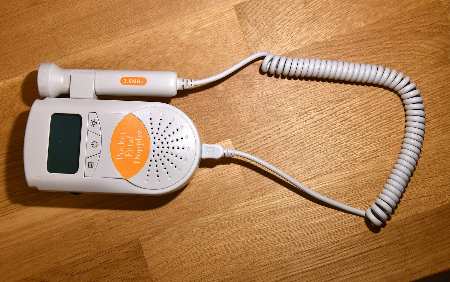
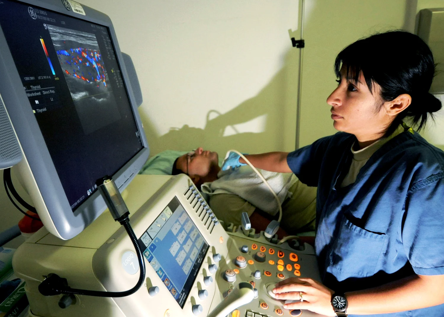
In diagnostic imaging, a transducer sends short pulses of high-frequency sound into the body and listens for echoes. Boundaries between tissues reflect some sound back. The system estimates depth from the time between sending a pulse and receiving its echo, then uses echo strength to build an image [2]. Because the process repeats quickly, the display can show moving structures and blood flow in real time.
A coupling gel reduces the air gap between probe and skin, allowing sound to pass into the body more effectively. Air reflects sound strongly, so even a thin air layer can obstruct transmission. The probe is positioned to answer a particular question, such as how a heart valve moves or where a needle is relative to a vessel. Imaging ultrasound is non-ionizing, unlike X-ray imaging, but it still introduces energy into tissue.
Different tasks, different safeguards
The cleaner's intended result is removal of soil from an instrument; the imaging system's result is information about anatomy or motion. Diagnostic ultrasound can slightly heat tissue and, in some circumstances, produce cavitation. The FDA describes it as generally considered safe when used prudently by appropriately trained providers, while recommending that exposure be minimized without losing diagnostic quality [2]. That is a different safety question from whether an instrument was adequately cleaned.
A useful mental experiment is to imagine placing a metal instrument in an imaging exam. The returning echoes might reveal an interface, but they would not establish that protein or debris had been removed. Conversely, putting a body part into a cleaning bath would not yield a clinically useful anatomical image. Shared wave physics does not make purpose, equipment, or procedure equivalent. The label matters less than what the device is designed to do and how its output is used.
Explore the physics: an explanatory diagram

Sources & further reading
- Cleaning CDC
- Ultrasound Imaging FDA
Prepared for the independent PSI Center reading library. About edition dates and source checking.
This explainer describes scientific principles. It does not assess individual treatment choices.