How small can a structure be before an image stops showing it as a separate feature? The answer depends not only on how many pixels fill the screen, but on how the scanner gathers signals and how strongly the structure differs from its surroundings. Resolution is the ability to tell two nearby details apart; visibility also depends on contrast, noise, motion, and the way an image is reconstructed.
Two dots, one blur
Imagine two tiny bright dots on dark paper. If they are far apart, each makes its own bright patch. Move them closer and the patches overlap until they look like one. That transition is a practical picture of spatial resolution: the smallest separation at which an imaging system can still distinguish neighboring features. A single feature’s apparent size and sharpness are related, but they are not identical to resolution.
Light itself places limits on optical imaging. Lenses form patterns rather than perfect points, and those patterns spread into neighboring areas. In tissue, light can also scatter or be absorbed before reaching a detector. Optical methods therefore cover different scales: microscopy can reveal cells and smaller structures, while methods such as optical coherence tomography (OCT) make cross-sectional views beneath surfaces such as the retina [1]. A large image on a monitor cannot recover detail the signal never carried.
A slice is not a pixel
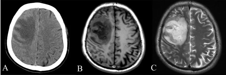
Computed tomography (CT) illustrates why resolution has more than one direction. A rotating X-ray source and opposing detectors collect measurements around the body; a computer reconstructs these into cross-sectional slices that can be stacked into a three-dimensional view [2]. Each slice represents a thickness of tissue, so a small object may be blurred or partly averaged with what lies above and below it. The display’s screen pixels are only the final grid; the slice thickness and the scanner’s measurements also constrain what can be separated.
Picture a narrow vessel crossing a stack of thicker slices. If it falls between slice centers, its signal is distributed across several images. It may appear faint, wider than it is, or absent if its contrast is weak. Reconstructing the stack in another orientation can help locate it, but does not create information beyond the collected data. CT’s ability to display slices successively or rotate a 3D reconstruction helps with localization, while the underlying sampling still sets boundaries [2].
Contrast decides what stands out
A feature can be physically large enough to resolve and still be difficult to see if it resembles the surrounding tissue. CT often shows dense bone clearly because it stops X-rays strongly, while soft tissues can have more similar X-ray responses. Contrast agents can make selected structures, such as blood vessels, more conspicuous by changing how strongly they attenuate X-rays [2]. In optical imaging, differences in absorption, scattering, reflection, or emitted light provide other kinds of contrast [1]. The method determines which differences become visible.
Noise makes the boundary less tidy. If the signal from a tiny feature is only slightly different from its background, random fluctuations can hide it. Increasing contrast may help, and combining imaging methods can add information: optical imaging, for example, can be paired with MRI to support monitoring of complex conditions [1]. But a sharper-looking image is not automatically a more accurate one. Detail must be supported by measured signal, and interpretation depends on the image’s limits.
What “visible” means
Resolution is often described as a distance, but clinical visibility is a joint result: size, separation, contrast, noise, motion, and reconstruction all matter. A bright, isolated speck can be seen when a dimmer feature of the same size disappears into its surroundings. Likewise, two features may each be visible but merge if they are too close together. These distinctions explain why there is no single smallest feature that every scan can show in every tissue.
A useful mental test is to ask what question an image can answer. Can it locate a structure, distinguish its boundary, or show two neighboring structures separately? Those are different demands. CT’s stacked slices can clarify where an abnormality lies, while optical methods can offer detail at scales suited to their particular interactions with tissue [2] [1]. Knowing which kind of visibility is needed is part of reading the image, not merely judging its apparent sharpness.
Sources & further reading
- Optical Imaging NIBIB
- Computed Tomography NIBIB
Prepared for the independent PSI Center reading library. About edition dates and source checking.
This explainer describes scientific principles. It does not assess individual treatment choices.