What can a wave tell us when it meets the boundary between two materials? A reflection marks a change: some property of the medium has shifted enough that part of the wave turns back. Medical imaging turns that return into evidence about hidden structure. Ultrasound listens for echoes, while optical methods measure returning light. The image is an inference from the reflected signal, not a direct view through a transparent body.
A reflection marks a change
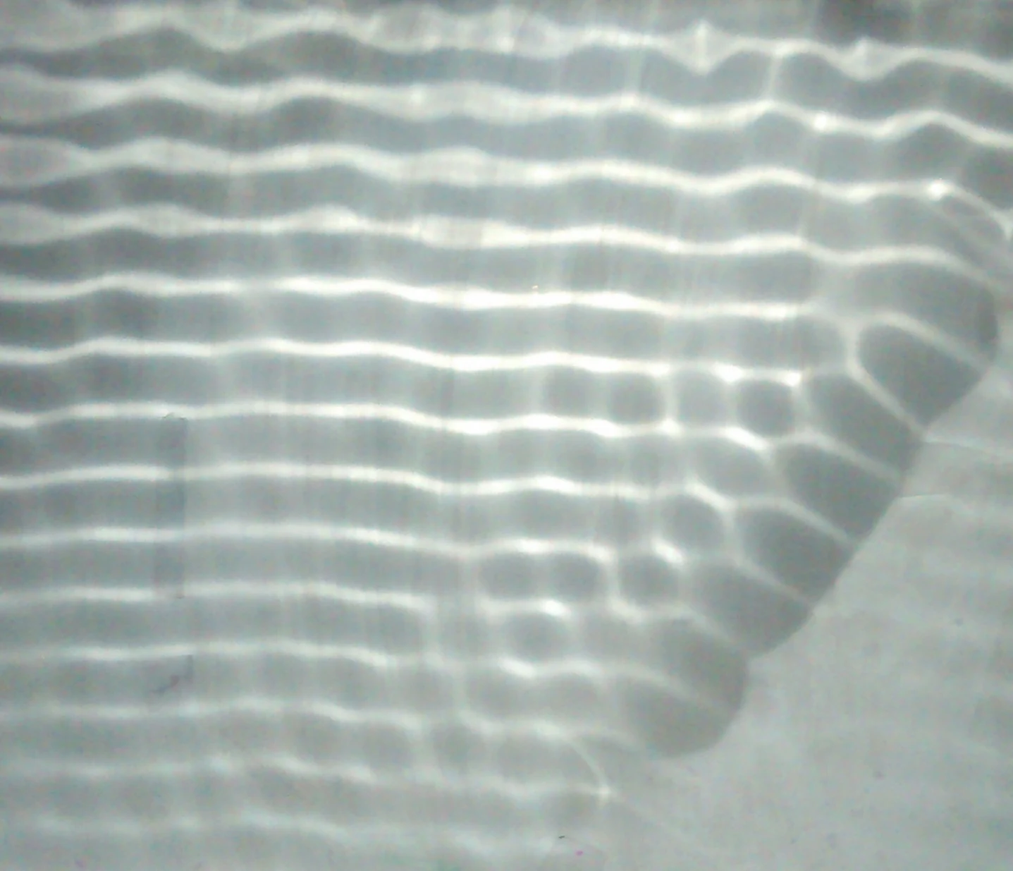
Imagine a ripple moving across a pond and reaching a floating mat of reeds. Some of the ripple continues; some returns. The division depends on how the wave interacts with the new material. In tissue, a boundary between fluid and soft tissue or between tissue and bone can reflect part of an ultrasound wave. The scanner detects the returning echo and uses its arrival time to estimate the boundary’s distance from the probe. [2]
The same broad idea applies to light: when light interacts with matter, it may be absorbed, scattered, reflected, or emitted. Those interactions can reveal biological structures at scales from molecules and cells to tissues and organs. A reflected signal is therefore information about a transition, not simply a miniature mirror image of the object. [1]
Turning echo time into depth
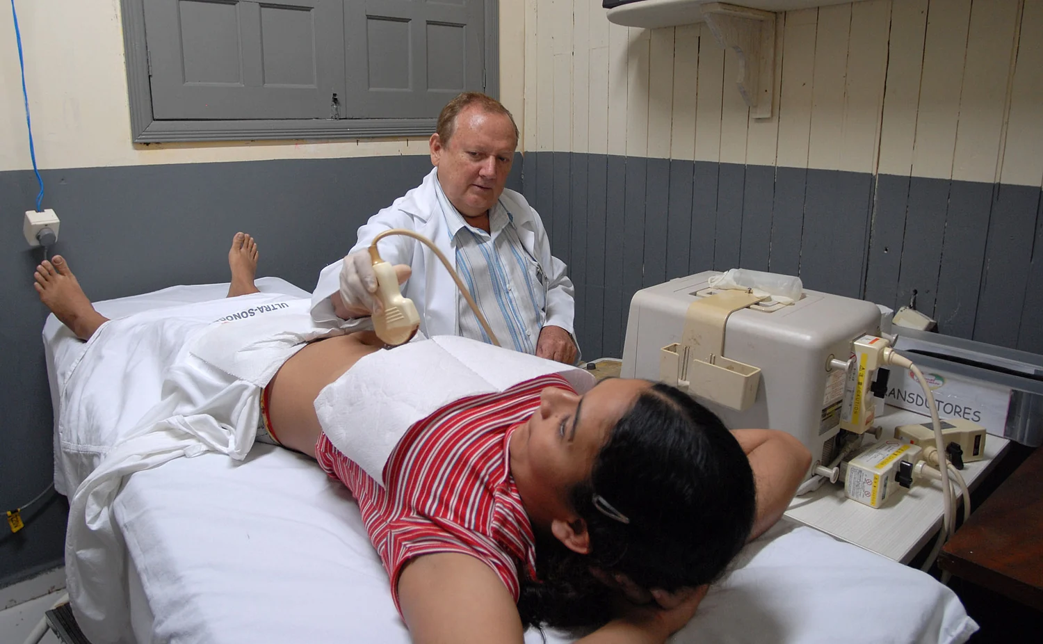
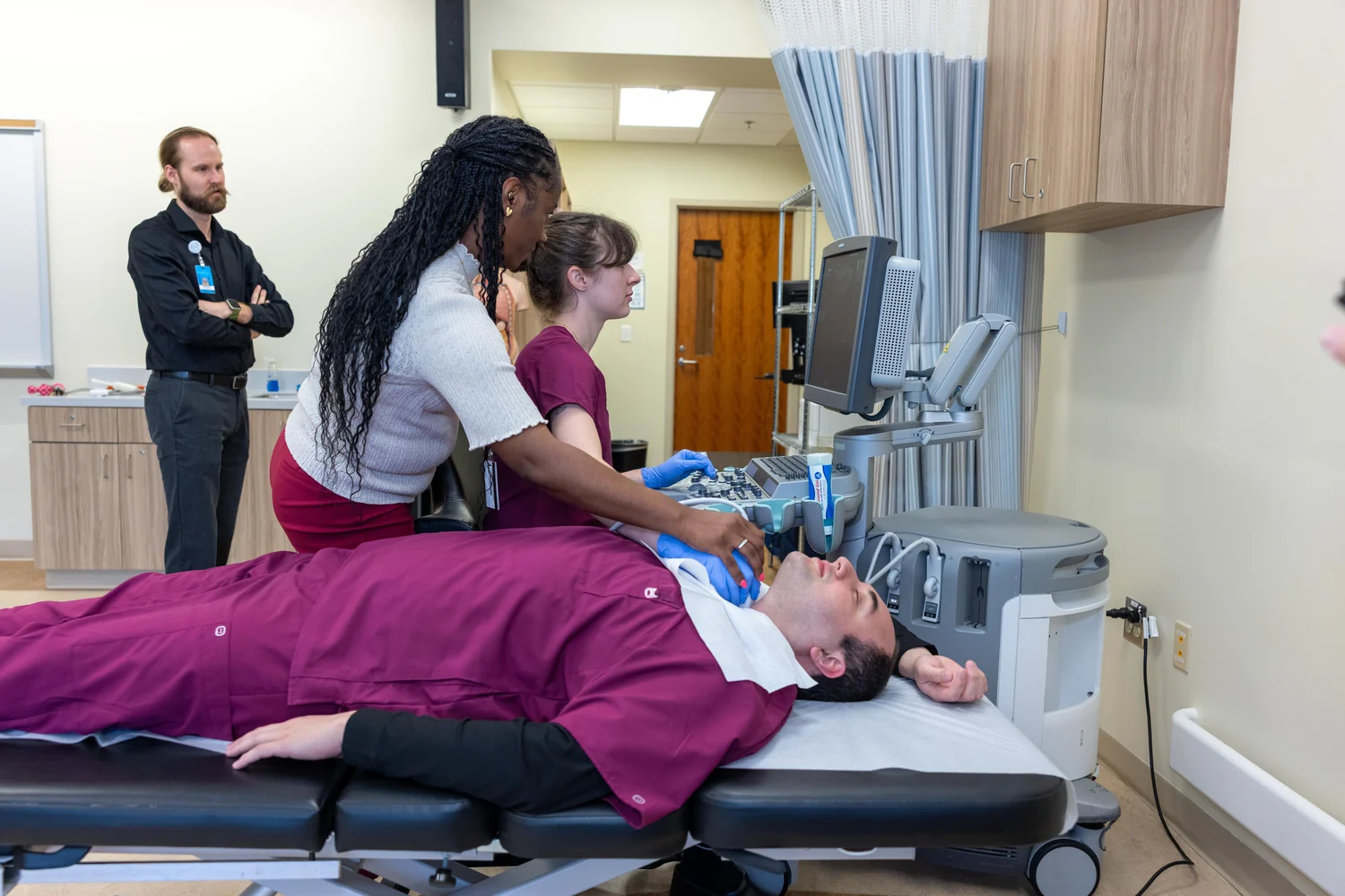
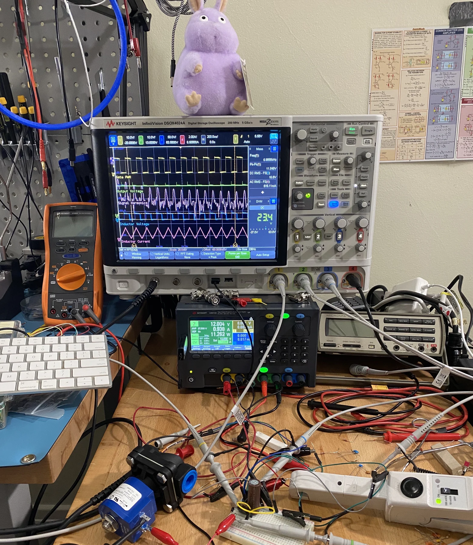
An ultrasound transducer both sends out sound and detects the echoes that come back. In many devices, piezoelectric materials convert electrical energy into sound and returning sound into electrical signals. If the scanner estimates the speed of sound in tissue, the time delay between sending and receiving helps calculate how far away a reflecting boundary lies. Repeating the measurement across directions yields a two-dimensional image. [2]
A simple thought experiment makes the timing logic clear. If two echoes return at different times, and sound speed is treated as known, the later one is assigned to a more distant boundary. The instrument does not need a camera to see the tissue directly; it builds a map from delay and signal strength. The assumptions matter, because the image depends on how well the chosen sound-speed model matches the tissues along the path. [2]
Light returns through a different route
Optical coherence tomography, or OCT, uses infrared light to obtain subsurface images. It is often compared with ultrasound because both can produce cross-sectional views from returning waves, but OCT relies on light rather than sound. The light’s interaction with tissue—including reflection and scattering—provides the information used to reconstruct layers. Clinicians use OCT to image structures within the retina, and it is also used in cardiology to help diagnose coronary artery disease. [1]
Optical imaging can exploit more than reflection alone. Different tissues absorb and scatter light in different ways, so measurements can reveal properties of soft tissue and changes associated with abnormal function. Other optical methods, such as photoacoustic imaging, use absorbed laser pulses to generate ultrasound waves that a transducer detects. That hybrid illustrates how a wave can be converted into another kind of signal before it is measured. [1]
What the return cannot tell by itself
A strong reflection can make a boundary easy to locate, but it can also limit what lies beyond it. Ultrasound does not image bone or air-filled tissues well under ordinary conditions because those materials interfere with sound transmission. Light, meanwhile, is scattered within tissue, which limits how far useful optical information can travel. In both cases, the returning signal is shaped by everything it encountered on the way in and out. [2] [1]
That is why a medical image is an interpretation constrained by the physics of the probe. A clinician must consider whether the relevant tissue was reachable, whether the signal was strong enough, and what features the method measures well. A reflection reveals that the wave met a change, but identifying the tissue and deciding what the change means requires context. The boundary is evidence; the image is a model built from that evidence. [2] [1]
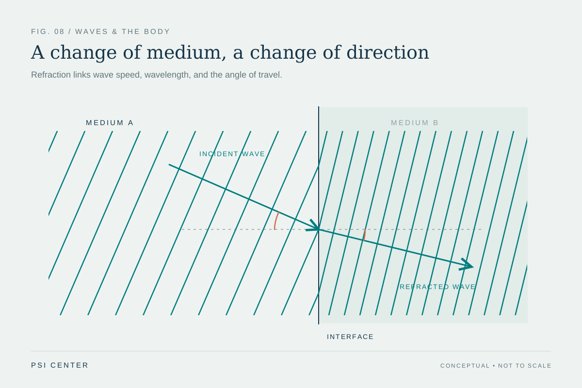
Explore the physics: an explanatory diagram
Sources & further reading
- Optical Imaging NIBIB
- Ultrasound NIBIB
Prepared for the independent PSI Center reading library. About edition dates and source checking.
This explainer describes scientific principles. It does not assess individual treatment choices.