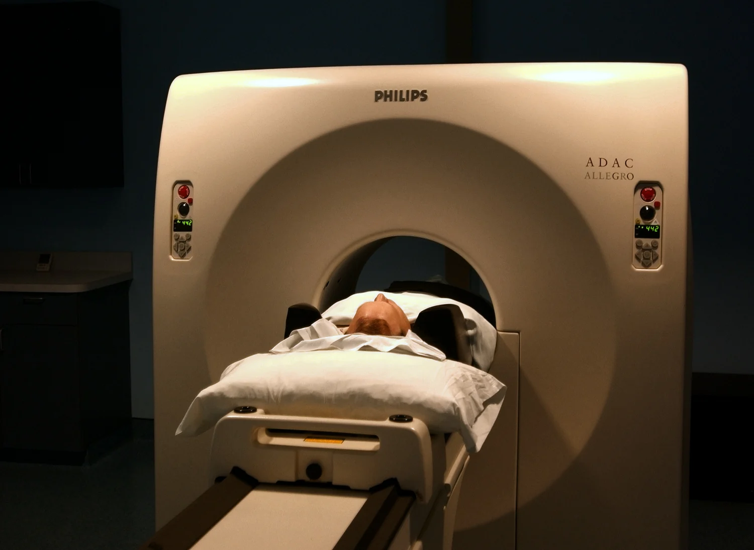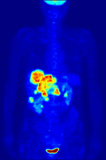
Why does a nuclear medicine scan happen at a particular time after a tracer is given? The answer begins with a tiny physical clock: radioactive decay. A tracer’s radioactive atoms emit detectable energy as they change, while the molecule carrying them travels through the body. The interval between administration and imaging matters because clinicians need enough tracer at the relevant destination, and enough signal left to measure it. The same timing principle informs some targeted treatments.
One atom, one unpredictable event
A radioactive atom has an unstable nucleus. It can change into a more stable arrangement by emitting particles or energy. For any one atom, the exact moment is unpredictable; for a large population, however, the fraction that remains radioactive follows a regular statistical pattern. The half-life is the time it takes for half of the radioactive atoms in a sample to decay. After another half-life, half of what remained decays, leaving one quarter of the original radioactive atoms.
That steady population pattern makes radioactive decay a clock, but not a timer that counts down to a single instant. If a sample contains many atoms, its activity—the rate of decays—falls predictably over time. In a patient, that physical decline happens alongside biological movement: the body distributes, binds, and clears the tracer. A scanner therefore detects the combined story of radioactive change and molecular traffic.
The carrier gives the signal a destination
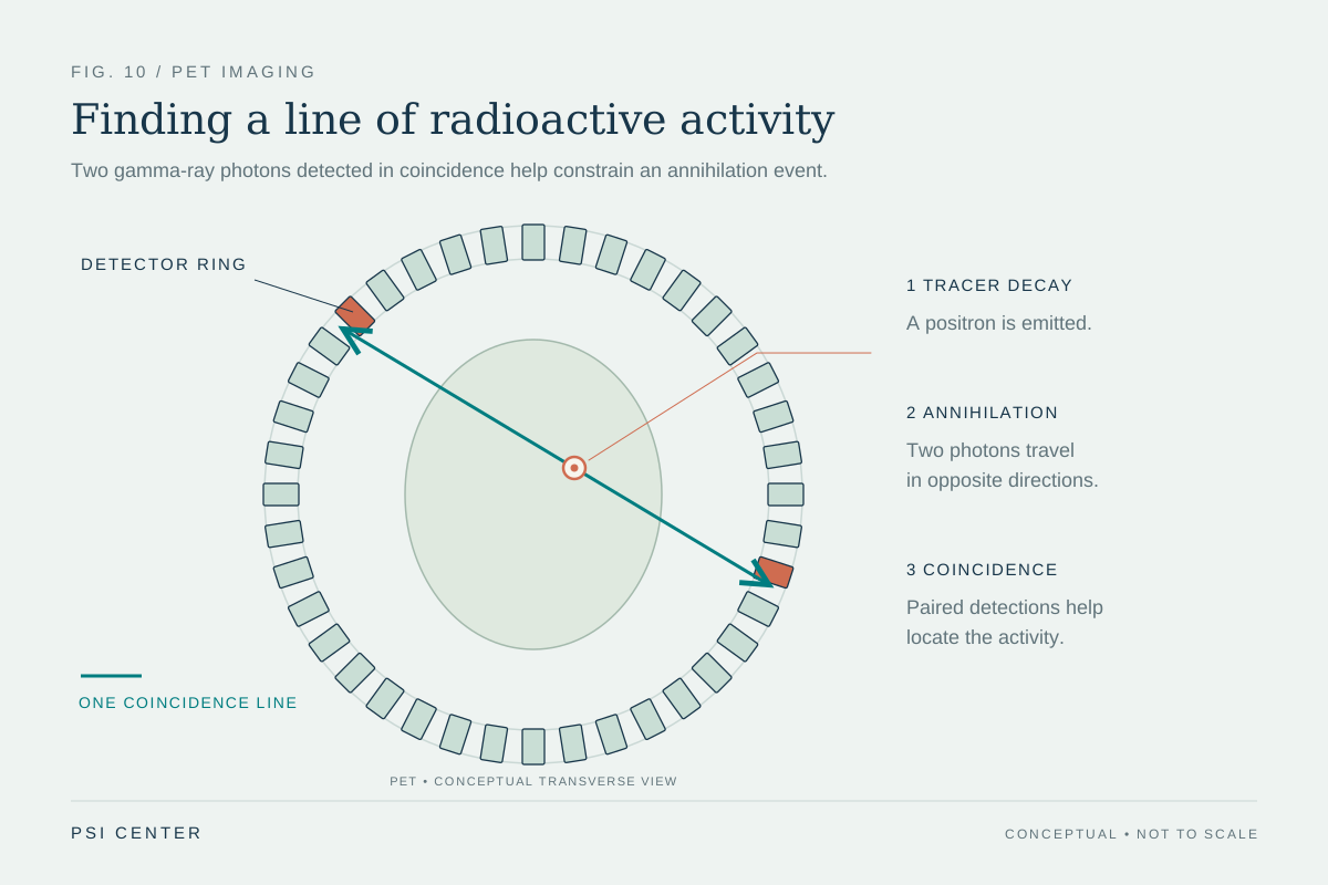
A radiotracer pairs a radioactive atom with a carrier molecule. The carrier helps determine where the tracer goes or what process it reflects. For example, a radiolabeled glucose-like molecule can accumulate in tissues that use glucose actively; PET cameras detect radiation emitted after positrons from the tracer meet electrons and annihilate, producing paired gamma photons [1]. The image maps signal, which clinicians interpret in light of anatomy and physiology.
This is why nuclear medicine is functional imaging: it can show patterns of molecular activity rather than only outlines of structures. SPECT detects gamma rays emitted directly by tracers, while PET detects the paired photons produced after positron decay [1]. In either case, the scanner must collect enough signal from the body to reconstruct an image. Waiting too long can mean fewer radioactive atoms remain; imaging too early may capture a distribution that has not yet become informative.
Choosing the imaging window
Imagine sprinkling glowing grains into a branching stream. The grains drift, some stick to particular surfaces, and their glow gradually fades. To learn which branch carries water toward a hidden pool, an observer must look after the grains have had time to travel but before most of their glow disappears. Nuclear medicine faces an analogous balance. The tracer’s chemistry, its biological route, and its half-life together shape the useful imaging window.
The administered tracer and the examination determine the details. Some radiopharmaceuticals are injected; others can be inhaled, swallowed, or delivered locally, depending on the process under study [1]. The aim is to use enough activity to provide useful information while limiting radiation exposure. The radiation dose from many diagnostic studies is comparable to routine x-ray or CT examinations, though each examination has its own purpose and dose considerations [1]. A shorter-lived isotope may reduce lingering activity, but it also places tighter demands on preparation and timing.
When the clock delivers treatment
In radiopharmaceutical therapy, decay is more than a source of diagnostic signal: the released energy can damage nearby cells. A targeting molecule can bind to a cancer cell, and radiation from its attached radioactive atom can injure that cell and, depending on the emitted radiation, nearby cells as well [2]. The carrier, isotope, and linker are selected as a system; the isotope’s decay properties influence how much energy is released and over what period.
Some treatment strategies use an imaging version of a molecule first to check whether it reaches a target, then use a related therapeutic version carrying a different radioactive atom [1]. This approach links molecular targeting to dosimetry, the calculation of radiation absorbed by tissues. It does not make treatment automatic: uptake varies, and surrounding organs may also receive radiation. Decay supplies a predictable physical clock, while biology determines where the clock is located and medicine determines how its signal or energy is used.
Explore the physics: an explanatory diagram
Sources & further reading
- Nuclear Medicine NIBIB
- Radiopharmaceuticals: Radiation Therapy Enters the Molecular Age National Cancer Institute
Prepared for the independent PSI Center reading library. About edition dates and source checking.
This explainer describes scientific principles. It does not assess individual treatment choices.