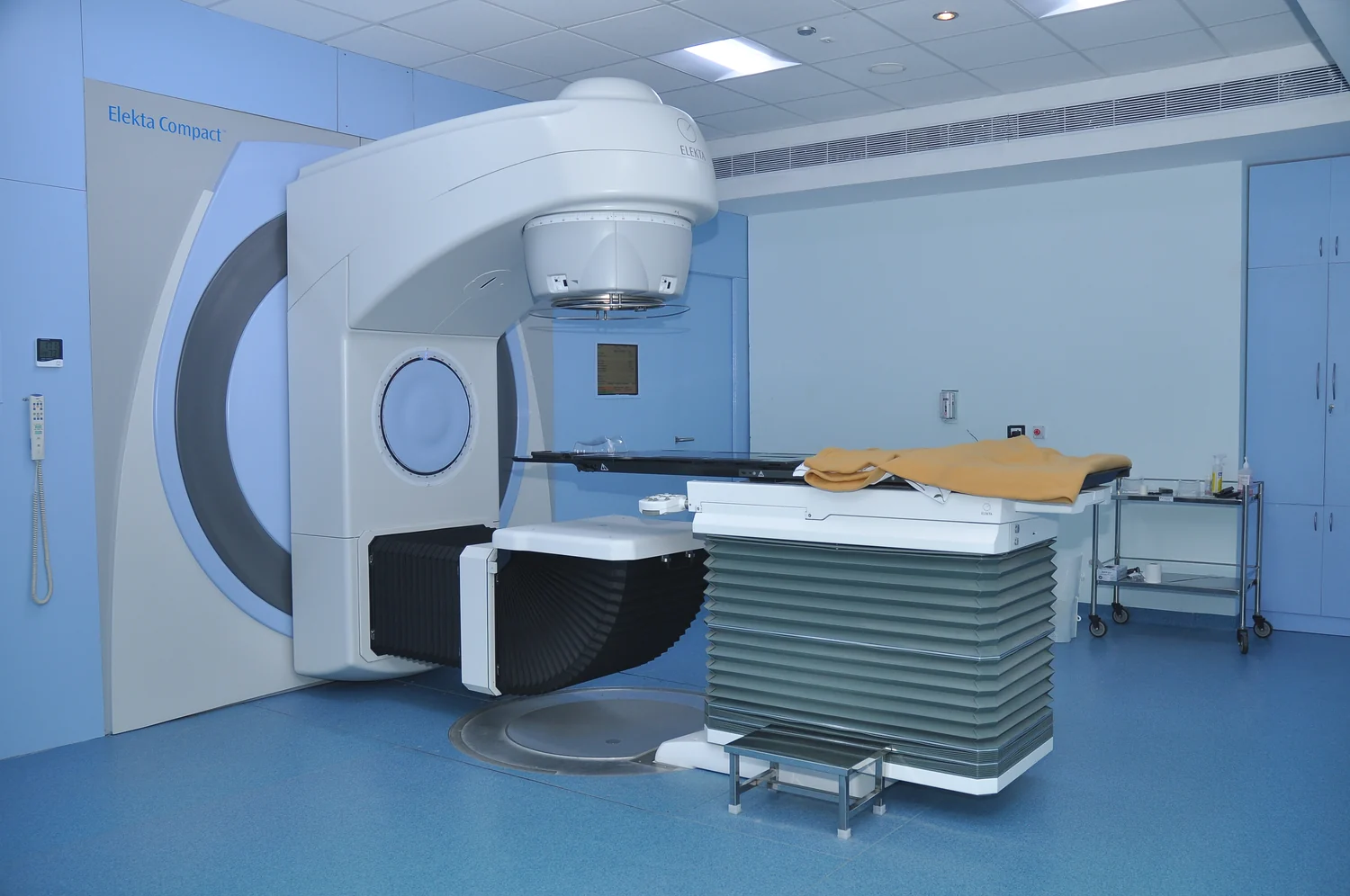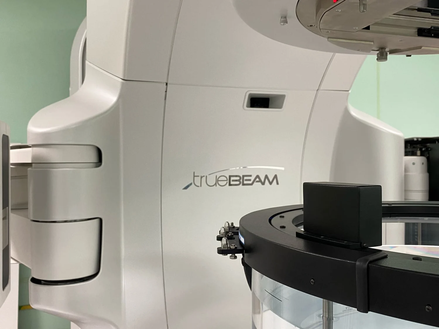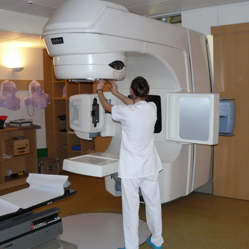
How can a radiation beam aimed from outside the body reach a tumor that cannot be seen from the treatment room? The answer begins with images and a repeatable position. Before treatment, clinicians use scans to map the target and plan the beam paths. On each visit, the patient is aligned with that plan, sometimes with new images to check whether anatomy has shifted. Image guidance connects a digital target to a real body lying on the treatment table.
First build a map
External beam radiation therapy sends radiation from a machine toward a specific part of the body. It is a local treatment: for a lung tumor, for example, the intended treatment area is the chest rather than the whole body. Planning images help define where radiation should go and what nearby normal tissue needs consideration. The aim is to deliver the prescribed dose to the tumor while limiting exposure to surrounding tissue. [1]
A planning session called simulation helps establish the treatment area and a consistent position. The team may use imaging and place small marks on the skin; a mold or mask can help limit movement. These are practical reference points, like aligning a map to landmarks before tracing a route. The image identifies anatomy, while the marks and positioning equipment help reproduce the geometry when treatment is delivered. [1]
From anatomy to coordinates
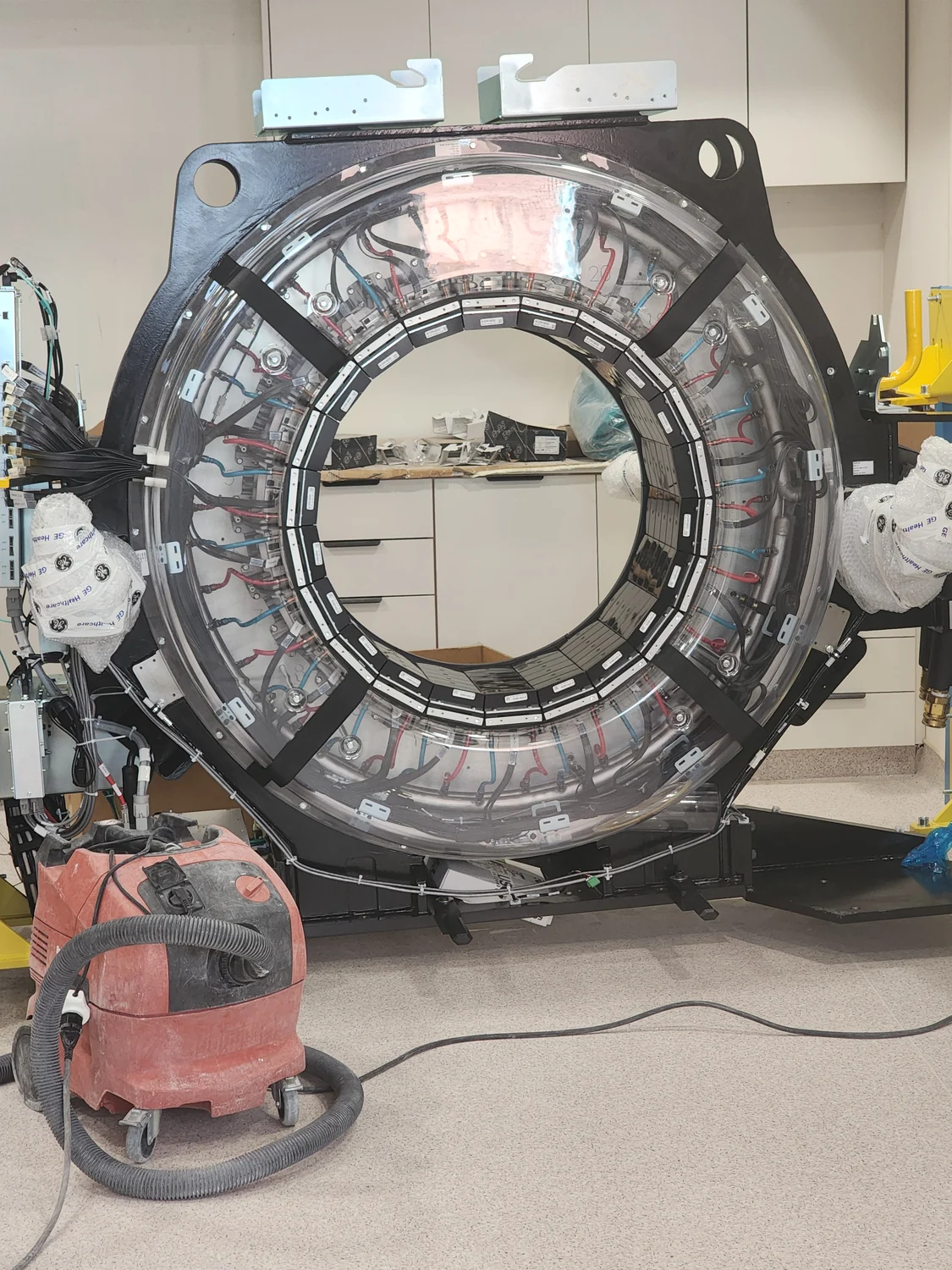
A CT scanner can produce cross-sectional images by rotating an X-ray source and detector around the patient. A computer reconstructs the measurements into slices, which can be stacked to show structures in three dimensions. Unlike one flat radiograph, the slices help a planner locate the tumor in relation to nearby organs and tissues. The scanner’s measurements are not the treatment beam itself; they provide a map used in planning. [2]
In three-dimensional conformal radiation therapy, computer analysis of CT, MRI, or PET images supports the design of beams shaped to the tumor. Intensity-modulated radiation therapy uses multiple smaller beams whose strengths can vary across the field. Different approaches shape dose in different ways, but all depend on knowing where the target and nearby structures are. A plan is a calculated route through anatomy, not a guarantee that anatomy will never move. [1]
Why check again at the session
A person’s position on one day may not match it exactly on another. Body shape, posture, or internal anatomy can change; tumors outside the brain may also move with ordinary processes such as breathing or digestion. Image-guided radiation therapy adds scans during treatment sessions, which computers process to detect changes in tumor size or location. If needed, position or dose can be adjusted to improve targeting and spare normal tissue. [1]
The concrete comparison is a flashlight aimed at a mark on a moving wall. A carefully measured angle is useful, but if the wall shifts, the spot no longer falls where intended. In treatment, imaging provides information about alignment and anatomy so staff can act on differences. It does not mean every treatment plan uses the same scans or adjustments; the technique depends on the treatment method and clinical plan. [1]
What imaging can and cannot settle
CT uses ionizing X-rays, and a CT examination involves more radiation than a conventional radiograph because the image is reconstructed from many projections. Its cross-sections can offer useful detail, including in complex bone anatomy, but imaging choices balance the clinical question with exposure. FDA guidance describes justification—whether the exam is needed—and optimization—using the lowest dose that gives adequate image quality—as central principles for exams that use ionizing radiation. [2] [1]
Image guidance is a tool for delivering a planned treatment with attention to where anatomy sits at the time. It cannot make radiation harmless, nor does a scan alone decide the appropriate treatment. Radiation beams can affect normal tissue along their path as well as the intended target; photons continue beyond a tumor, while proton beams stop once they reach it, according to the NCI description. Planning and verification are ways to manage geometry and dose, not claims that one beam type or imaging method is best for every case. [1]
Sources & further reading
- External Beam Radiation Therapy National Cancer Institute
- Computed Tomography NIBIB
Prepared for the independent PSI Center reading library. About edition dates and source checking.
This explainer describes scientific principles. It does not assess individual treatment choices.