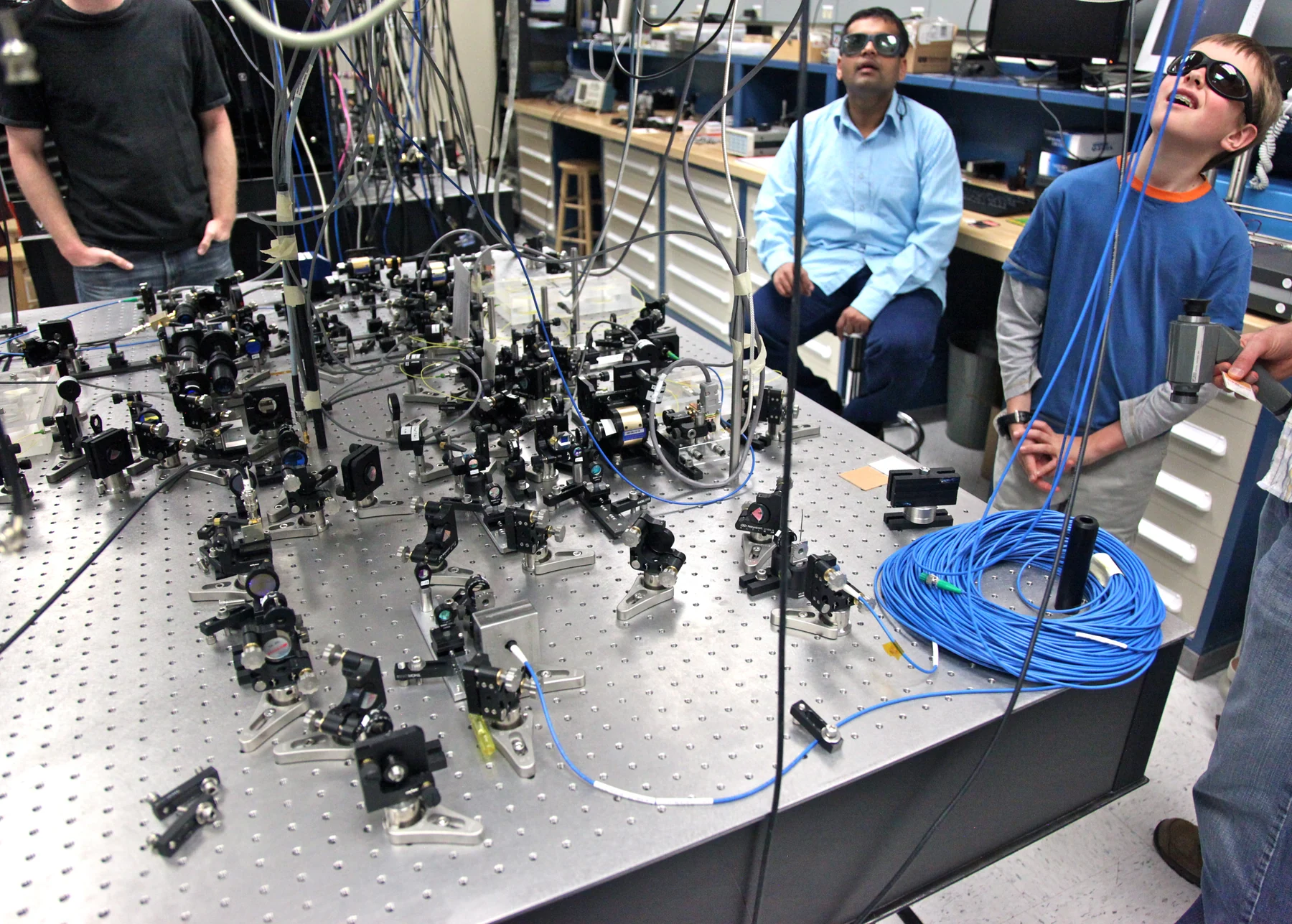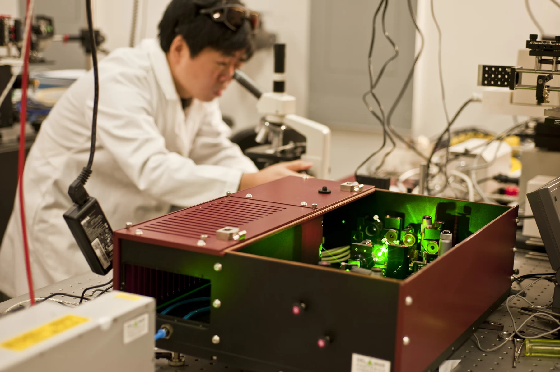
How can a beam of light both cut tissue and help stop bleeding? A laser concentrates light into a narrow region, and tissue absorbs some of that energy. The absorbed energy can become heat, changing proteins and cells; with suitable delivery, that change may remove tissue or seal small vessels. The outcome depends on wavelength, target absorption, intensity, exposure time and heat flow into nearby tissue—not on a laser having a single universal effect.
Light becomes heat where it is absorbed
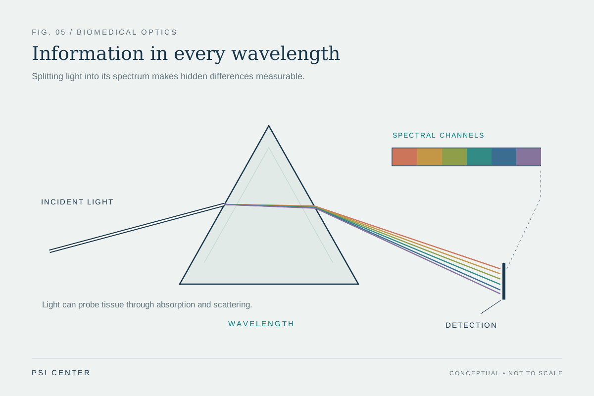
A laser produces an intense, narrow beam of light. Light carries energy, but tissue does not absorb every wavelength equally. Some wavelengths are absorbed more strongly by particular components, while others travel farther before depositing much of their energy. Selecting a wavelength therefore helps determine where energy is delivered and which tissue features are affected. In cancer care, laser therapy can target tissue by choosing light that tumor cells absorb differently from nearby normal cells [1].
The path from beam to biological change has several steps: light enters tissue, molecules absorb some of it, and that absorbed energy can raise local temperature. If enough heat accumulates, proteins change shape and cells are damaged. A useful mental experiment is to compare a brief, intense pulse with a weaker beam held longer. Both can deliver energy, but the time available for heat to diffuse differs, so the heated region and tissue response can differ too.
Cutting and coagulation are related effects
A surgical laser can remove or destroy tissue. In some procedures, the heat also seals the cut and helps reduce bleeding. NCI describes lasers used to seal blood vessels during surgery, as well as nerve endings and lymph vessels in particular settings [1]. These effects are connected because both involve energy deposition and tissue heating, but a cut and a seal are not identical goals: one removes or divides tissue, while the other aims to close a small vessel or structure.
How well this works depends on placing and controlling the beam. Some lasers can cut near the surface without penetrating as deeply; others are delivered through optical fibers in an endoscope to reach internal sites [1]. A fiber is a thin strand that transmits light to a selected location. Narrow delivery can help concentrate treatment, but it does not mean surrounding tissue is automatically untouched. Heat can spread beyond the directly illuminated spot, and the clinician must account for that when aiming and applying the energy.
When the goal is a heated region
Laser interstitial thermal therapy, or LITT, is a form of localized heating in which laser light is delivered through a fiber to tissue. It belongs to the broader family of hyperthermia approaches, which use heat to damage cancer cells and are generally used alongside other treatments rather than as a stand-alone promise of a cure [1] [2]. The shared physical idea is raising temperature in a target region; the specific delivery method and intended effect differ by treatment.
Heat is a physical dose spread across space and time. A probe or fiber can place energy in a small region, but tissue conducts heat away from the source. NCI describes monitoring temperatures in and near tumors during some hyperthermia procedures, illustrating why both target and neighboring tissue matter [2]. Excess heating can injure normal tissue, while insufficient heating may not achieve the intended tissue effect. The desired balance depends on the procedure and its clinical context.
A tool with limits and a setting
Lasers are used in selected cancer treatments, including for some surface cancers and precancers, and can be used through endoscopes for internal sites [1]. Their capacity to cut, destroy or help seal tissue does not make every tumor suitable for laser treatment. Access to the target, the type and location of tissue, equipment and specialized training all shape whether a laser is appropriate. NCI also notes practical drawbacks such as cost, specialized equipment and the possibility that treatment effects may not last.
Hyperthermia more broadly is not widely available and is studied or used in particular settings, often with other cancer therapies [2]. These distinctions matter: a laser is a way to deliver light, LITT is a laser-based heating method, and hyperthermia describes treatment using heat. Explaining how energy can create a tissue effect is not evidence that a particular treatment improves outcomes for every patient. The physics clarifies what the beam can do; clinical evidence and the intended use determine what claims are justified.
Explore the physics: an explanatory diagram
Sources & further reading
- Lasers in Cancer Treatment National Cancer Institute
- Hyperthermia to Treat Cancer National Cancer Institute
Prepared for the independent PSI Center reading library. About edition dates and source checking.
This explainer describes scientific principles. It does not assess individual treatment choices.