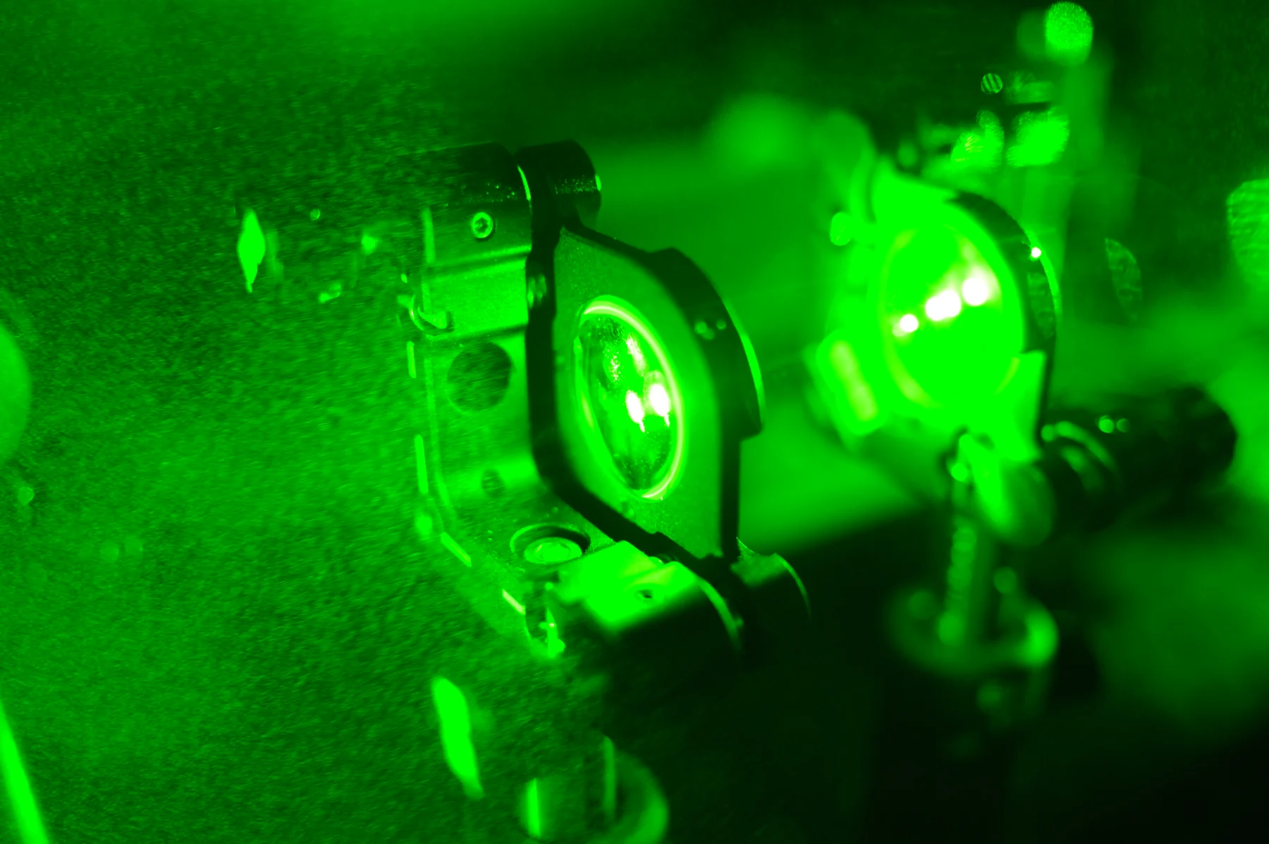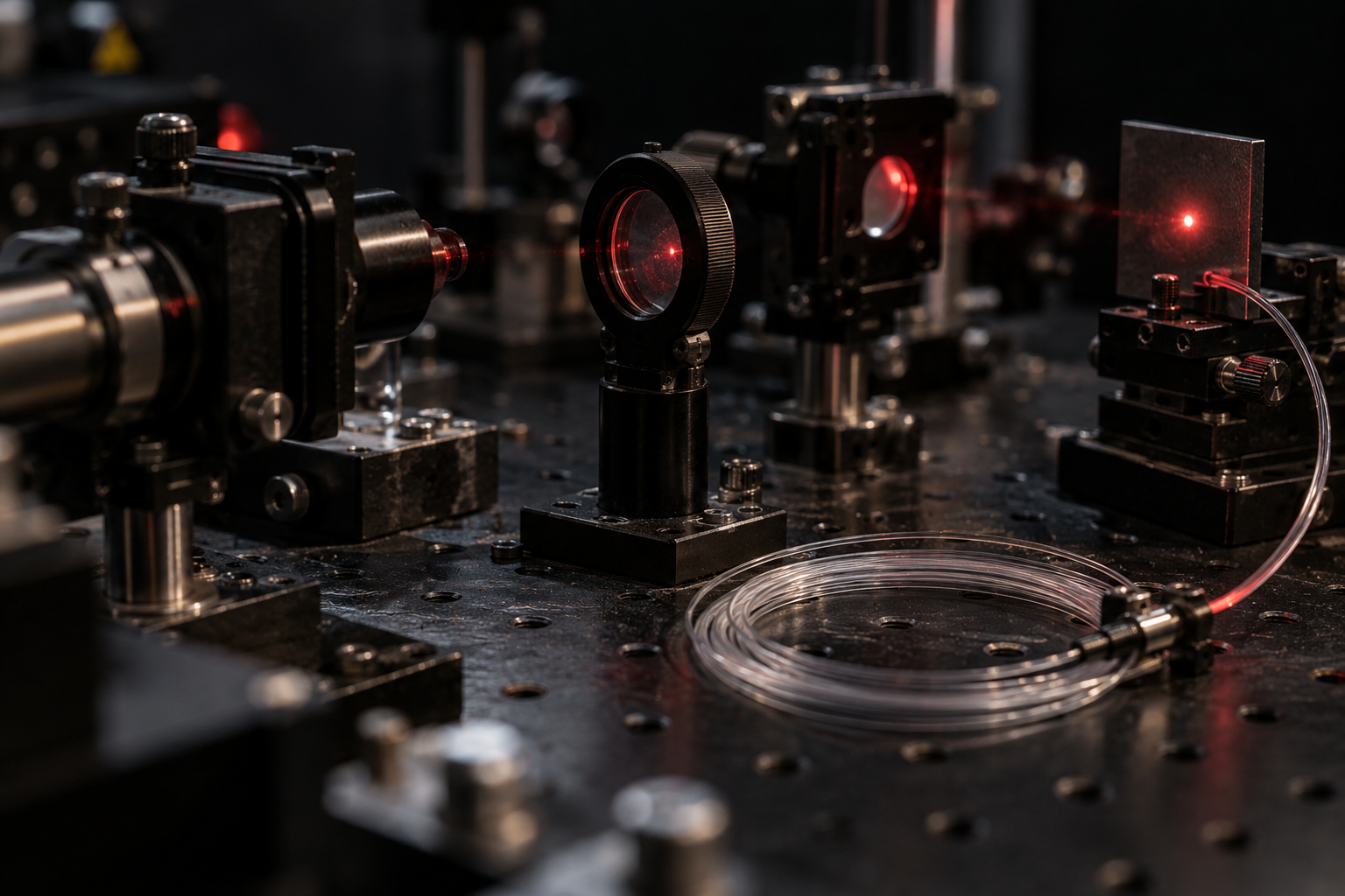
How can light reach a small patch of tissue deep inside the body without opening a large path for it? One answer is a flexible optical fiber: a strand that guides light along its length and can travel through a narrow instrument. It turns light from a source outside the body into a tool delivered at a selected site. The same route can support imaging or treatment, but those jobs use light in different ways.
Keeping light inside a strand
An optical fiber has a central core surrounded by material that helps confine light. At the boundary, light can reflect back into the core rather than escaping. With the geometry and materials chosen for the fiber, repeated internal reflections guide the beam along the strand, including around gentle bends. A fiber is not a tiny flashlight; it is a controlled path that transports light from one end to another.
That path matters because light normally travels in straight lines through open space. A narrow flexible fiber can pass through an endoscope, a slender tube used to reach internal tissues through a natural opening. The endoscope can carry fibers for illumination, optical imaging, or therapeutic light. The clinician can view a cavity and direct light at a local target without requiring the beam to cross the body from the outside [2].
Seeing with returned light
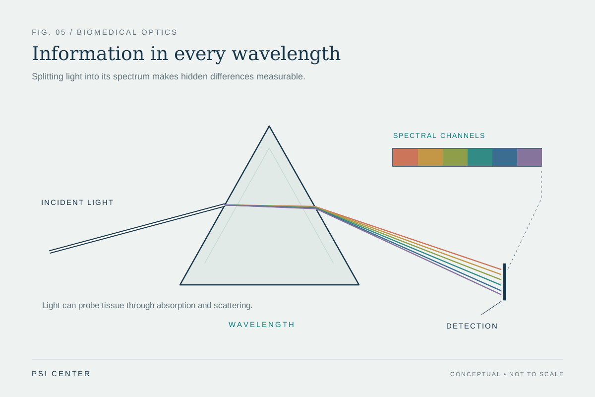
Imaging depends on light interacting with tissue. Some wavelengths are absorbed, some scattered, some reflected, and some emitted. Instruments collect selected portions of that light and form an image or map. In endoscopy, fibers and miniature cameras bring optical methods into internal cavities such as the stomach, allowing tissue to be viewed in its natural setting [1].
Different methods extract different information. Optical coherence tomography, or OCT, uses infrared light to make cross-sectional views of structures below a surface; it is used to examine the retina and can also support assessment of coronary arteries. Photoacoustic imaging starts with laser pulses absorbed by tissue; the resulting ultrasound waves are detected to create images. These examples show that “medical light” is not a single technique: the wavelength, tissue interaction, detector, and desired information all matter [1].
Delivering local energy
For laser therapy, the fiber can carry an intense, narrow beam to a specific site. Laser light can cut or destroy selected tissue, and it can seal small blood vessels during a procedure. The energy is local: it is aimed at a particular part of the body, rather than treating the whole body. In some uses, lasers travel through an endoscope to reach internal organs; the beam is then directed at the intended tissue [2].
Wavelength selection matters because different tissues absorb different colors of light. If a target absorbs a chosen wavelength more strongly than nearby tissue, the energy can be concentrated more selectively. Absorbed light becomes energy in the tissue, often heat, which can alter or destroy cells. This does not mean that a laser automatically distinguishes every diseased cell from every healthy one. The operator, instrument, target, and treatment plan determine what the beam reaches and what effect it has [2].
One route, different tasks
A fiber’s shared role in imaging and therapy can invite confusion. For imaging, light is sent in or collected from tissue to reveal structure or properties. For treatment, a laser delivers enough energy to alter tissue. The fiber is the delivery route; the purpose comes from the rest of the system. A light-guiding strand does not itself diagnose disease or guarantee that a treatment will spare surrounding tissue.
Lasers can have practical advantages in selected procedures, including precise local access and the ability to seal vessels while cutting. They also require specialized equipment and trained operators, and some effects may need repeating [2]. Meanwhile, optical imaging can be combined with other methods because each measures different physical properties. An image may map absorption or scattering; a treatment beam may deposit energy. Keeping those roles distinct makes the technology easier to understand: fibers carry light, while the clinical task depends on what the light is designed to measure or do [1].
Explore the physics: an explanatory diagram
Sources & further reading
- Optical Imaging NIBIB
- Lasers in Cancer Treatment National Cancer Institute
Prepared for the independent PSI Center reading library. About edition dates and source checking.
This explainer describes scientific principles. It does not assess individual treatment choices.