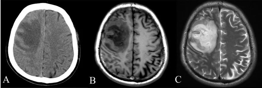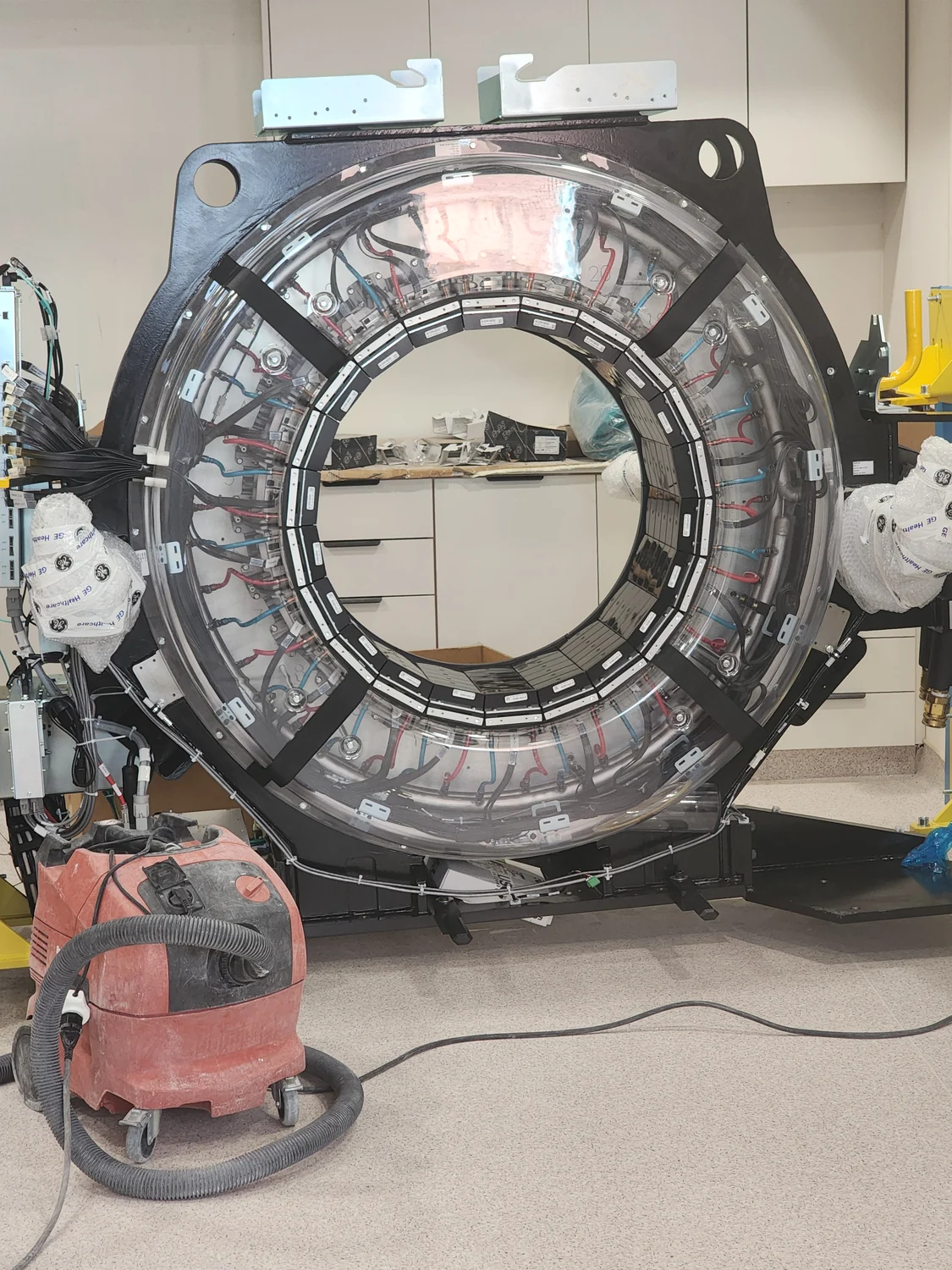
Why can two scans of the same body look different even when neither scanner is broken? An image is not a direct photograph of internal anatomy. It is a reconstruction from measurements, shaped by the physics of the scan and by choices made in computation. What appears as noise may be a normal consequence of collecting finite data, while a fault is only one possible cause.
A scan begins with measurements
Computed tomography (CT) provides a clear example. An X-ray source and detector rotate around the patient, recording how much radiation passes through the body from many directions. A computer uses those snapshots to reconstruct cross-sectional images [1]. The displayed slice is therefore calculated from measurements; it is not a picture captured by a camera sitting inside the body.
Measurements have limits. A detector registers a finite number of X-rays, and the signal can fluctuate. When fewer useful measurements are available, random variation may be more visible in the reconstructed image. That variation is often called noise. It can resemble grain in a photograph taken in dim light, where the camera receives less information about the scene.
Reconstruction shapes what we see
To turn projections into slices, software combines information gathered at different angles. The reconstruction method affects the balance between visible detail, smoothness, and noise. A model is a mathematical representation used to simulate or interpret a complex system. Computational modeling can draw on physical principles, observed data, or a mixture of both [2]. In imaging, reconstruction algorithms use assumptions about how measurements relate to structures inside the body.
Imagine trying to infer the shape of a mug from shadows cast on a wall. More angles provide more clues, but the result still depends on how the clues are combined. If the model is mismatched to the situation, artifacts can appear; if the data are sparse or noisy, genuine boundaries may become uncertain. A streak or speckle does not, by itself, identify a faulty component.
Noise, blur, and artifacts differ
Noise is random variation that can obscure detail. Blur softens edges, often because of motion or limitations in the system’s ability to resolve small structures. An artifact is a pattern that does not correspond faithfully to anatomy, such as streaks caused by particular interactions between the X-rays and the scanned body. These effects can overlap visually, but they arise through different mechanisms and may call for different responses.
For CT, the amount of radiation used influences how many detected photons contribute to the image. Reducing exposure can reduce risk, but it can also increase noise if too few photons reach the detector. The FDA advises imaging teams to use protocols that provide the lowest radiation dose that still yields image quality adequate for diagnosis or intervention [1]. The goal is not maximum sharpness at any cost; it is information fit for the clinical task.
A visual defect needs an explanation
A scan’s appearance also depends on patient movement, body size, positioning, contrast material, and the body region being examined. A moving patient can blur boundaries even if the equipment is functioning correctly. Dense structures can alter the path of X-rays and complicate reconstruction. In other forms of imaging, different physical signals create different limitations; the word “noise” does not name one universal failure mode.
Quality control checks help teams identify equipment problems, but image interpretation considers the acquisition and processing conditions too. Computational models can test how systems behave under different assumptions, and biomedical research uses them to explore complex processes [2]. In routine imaging, the practical question is whether the resulting image answers the medical question at adequate quality. A noisy image may be usable, while a clean-looking one may still omit important information.
Sources & further reading
Prepared for the independent PSI Center reading library. About edition dates and source checking.
This explainer describes scientific principles. It does not assess individual treatment choices.