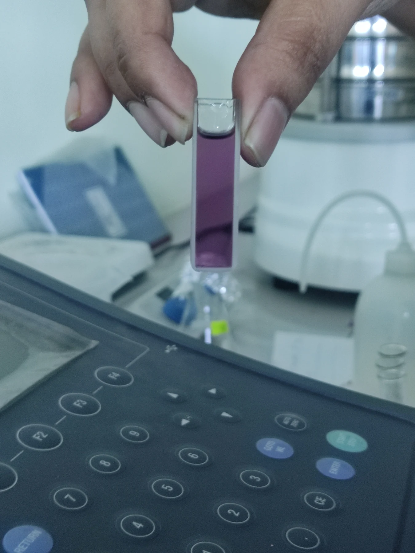
How can a clip on a finger estimate oxygen in blood without drawing any blood? A pulse oximeter shines red and near-infrared light through tissue and compares how much returns or reaches a detector. The device does not directly count oxygen molecules; it infers a value from light absorption and a calibration relationship. That distinction explains why calibration matters—and why a neat digital reading is not the same as an unquestionable measurement.
Blood changes the light that gets through
Oxygenated and deoxygenated hemoglobin absorb red and near-infrared light differently. A fingertip device emits both wavelengths and measures the detected light. Because the heartbeat changes the amount of arterial blood in the finger, the instrument can isolate a pulsating component from other tissues and use a ratio of the signals to estimate oxygen saturation [1].
Imagine looking through two filters that change their darkness as a pulse of blood passes beneath them. The device’s challenge is to infer what fraction of hemoglobin carries oxygen from the different absorption patterns. It is an indirect measurement: skin, bone, motion, blood flow, and the exact optical path can affect the signal. That is why a sensor needs a validated mapping between measured light and the saturation value it displays, rather than relying on a simple raw brightness reading [1].
Calibration ties a reading to references
In measurement science, calibration compares an instrument’s response with known references and establishes how its outputs relate to those reference values. Metrological traceability means a documented, unbroken chain of calibrations connects a measurement result to specified standards, with each link contributing uncertainty [2]. For a medical sensor, calibration is part of making its inference interpretable: it helps establish what a signal corresponds to under defined conditions.
Traceability does not guarantee that a measurement is fit for every purpose. NIST emphasizes that the uncertainty must be small enough for the particular measurement need [2]. A scale calibrated for a narrow range, for instance, may not be appropriate for a different range or setting. Likewise, a pulse oximeter’s calibration cannot by itself ensure accuracy for every person, oxygen level, or circumstance. Calibration is a chain of evidence about measurement, not a promise that all sources of error disappear.
Real fingers complicate the optical model
Blood flow in the fingertip can be reduced by cold, constricted vessels, blood loss, or some health conditions. Nail polish may interfere, and movement can create motion artifact—a disturbance in the signal that is not caused by a true change in oxygenation. Accuracy may also decline at lower oxygen saturation, just when a dependable estimate can matter most [1]. These are not merely software problems: the sensor is trying to interpret light after it has passed through a changing biological material.
Skin pigmentation is another important factor. Melanin absorbs across wavelengths that include red and near-infrared light. NHLBI describes evidence that pulse oximeters can overestimate oxygen saturation in patients with darker skin tones, potentially contributing to delayed recognition of low oxygen [1]. A calibrated instrument may still perform unevenly if its calibration data or design does not adequately account for the range of people and conditions in which it is used.
Better calibration requires representative conditions
Researchers have explored alternative optical approaches, including green-light devices that measure reflected light rather than transmission through the finger, and calibration using a skin-pigment scale. These are research efforts aimed at addressing particular sources of error; they should not be confused with a guarantee that any one newer design solves every limitation [1]. A device can change its wavelength, geometry, or model and still need testing against suitable reference measurements across relevant users and conditions.
The key idea is that an oxygen reading is an estimate supported by a chain: light interacts with tissue, a detector captures a signal, a model converts it, and calibration relates that result to references. Each step has limits. When clinicians interpret a reading, they also consider the person and the measurement conditions; no single number carries all that context. Calibration makes the number more meaningful, while diverse testing and careful interpretation address what calibration alone cannot [2] [1].
Sources & further reading
Prepared for the independent PSI Center reading library. About edition dates and source checking.
This explainer describes scientific principles. It does not assess individual treatment choices.